| This is an old, no longer maintained version of the website, for new one please visit: http://gsn.io.gliwice.pl |

 |

Gliwice Scientific Meetings 2013 ~~~ Poster abstracts ~~~
Abstracts are ordered according to nine main themes. Number next to the abstract title correlates with poster nu
Cancer (Cell) Biology 1. ANALYSIS OF CDK1 GENE EXPRESSION IN SQUAMOUS CELL CARCINOMA OF LARYNX
Kinga Bednarek1, Magdalena Kostrzewska-Poczekaj1, Marcin Szaumkessel1, Maciej Giefing1, Katarzyna Kiwerska1, Reidar Grenman2, Krzysztof Szyfter1,3, Małgorzata Jarmuż Szymczak1,4
1Institute
of Human Genetics, Polish Academy of Sciences, Poznan, Poland; 2Department
Laryngeal squamous cell carcinoma (LSCC) is one of the most common head and neck cancers. Because of difficulties in diagnosis and treatment, 5-year survival rate remains low, despite development of chemo- and radiotherapy. The attention was focused on head and neck cancer genetics – searching for new oncogenes. Aim of the research was the analysis of CDK1 gene. As a first step we analyzed larynx squamous cell carcinoma cell lines, and next we used material derived from primary larynx tumor samples from patients admitted to Poznan University of Medical Sciences. The study included both analyses of gene expression and an attempt to explain the mechanism that can be responsible for the observed changes. We applied techniques that involved DNA (pyrosequencing, microarray-based DNA-copy number analysis), RNA (microarray-based gene expression analysis, real-time PCR), microRNA (microarray-based microRNA expression analysis), proteins (Western Blot). We have shown that expression for CDK1 - on mRNA and protein level – is increased as compared to non-cancer control derived from larynx (total RNA and larynx tissue whole cell lysate). We found that these changes are not connected with gene amplification or duplication. We also observed that DNA methylation level and microRNA expression are on an equal level in analyzed material and non-cancer controls. Further analysis of mechanism for the observed changes explanation is still in progress. Our results indicate different expression profile in LSCC cell lines and larynx tumors, and we propose CDK1 gene as a new potential oncogene in LSCC. 2. ANALYSIS OF THE MONOCYTE CHEMOTACTiC PROTEIN-1-INDUCED PROTEIN 1 (mcpip1) GENE EXPRESSION IN HUMAN NEUROBLASTOMA
Elżbieta Boratyn1, Irena Horwacik1, Anna Skalniak1, Sylwia Tyrkalska1, Małgorzata Durbas1, Maria Łastowska2, Barbara Lipert3, Jolanta Jura3, Hanna Rokita1
1Laboratory of Molecular Genetics and Virology, Faculty of Biochemistry, Biophysics and Biotechnology, Jagiellonian University, Gronostajowa 7, 30-387 Kraków, Poland; 2Department of Pathology, Institute “Pomnik – Centrum Zdrowia Dziecka”, Aleja Dzieci Polskich 20, 04-730 Warszawa, Poland; 3Department of General Biochemistry, Faculty of Biochemistry, Biophysics and Biotechnology, Jagiellonian University, Gronostajowa 7, 30-387 Kraków, Poland.
The recently discovered MCPIP1 (monocyte chemotactic protein-1-induced protein 1) multidomain protein encoded by the MCPIP1 (ZC3H12A) gene, has so far been described as a new transcription [1] and differentiation [2] factor, a ribonuclease [3, 4], and deubiquitinase [5]. However, its role in cancer development is not yet established. Neuroblastoma (NB) is an embryonal malignant neoplasm of the postganglionic sympathetic nervous system, exhibiting considerable clinical and biological heterogeneity. It is the most common extracranial solid tumour in children, representing approximately 10% cancers in infancy and childhood. The main reason of the low cure rate of children with high risk neuroblastoma is resistance of cancer cells to treatment. Attempts to improve the outcome of advanced neuroblastoma have focused so far mainly on intensification of the induction and consolidation phases of chemo-radio therapy, with or without stem cell rescue [6]. We studied MCPIP1 gene expression in several cell lines like BE(2)-C, IMR-32, HTLA230 and SH-SY5Y. It was observed that the low expression of the MCPIP1 protein measured in human neuroblastoma cell lines might be important for their survival. Enforced MCPIP1 gene expression in BE(2)-C cells performed through transfection of plasmid constructs bearing either MCPIPwt or a mutant form lacking the RNase domain, caused significant decrease in neuroblastoma cell proliferation and viability. Additionally, analysis of microarrays data for MCPIP1 mRNA expression levels in human primary neuroblastoma tumours showed a lack of expression of the MCPIP1 transcript in all primary tumours. Transcripts of several genes known to be involved in MCPIP1 regulation, like MCP1 (CCL2), MCP-1 receptor (CCR2) and IL-1β receptor – IL1R1, as well as NFκB family members were also evaluated on the same set of primary neuroblastoma tumours. These results may help to gain more insight into the MCPIP1 gene expression levels importance in cancer development and indicate the possible signalling pathways involved in neuroblastoma cell survival.
References: [1] Zhou L., Azfer A., Niu J., et al. (2006) Monocyte Chemoattractant Protein-1 induces a novel transcription factor that causes cardiac monocyte apoptosis and ventricular dysfunction Circ. Res. 98, 1177-1158. [2] Vrotsos E.G., Kalattukudy P.E., Sugaya K. (2009) MCP-1 involvement
in glial differentiation [3] Matsushita K., Takeuchi O., Standley D.M., et al. (2009) Zc3k12a is an RNase essential for controlling immune responses by regulating mRNA decay Nature 458, 1185-1190. [4] Mizgalska D., Węgrzyn P., Murzyn K., et al. (2009) Interleukin-1-inductible MCPIP protein has structural and functional properties of RNase participating in degradation ol IL-1-mRNA FEBS J. 276, 7386-7399. [5] Liang J., Saad Y., Lei T., et al. (2010) MCP-iduced protein 1 deubiquitinates TRAF proteins and negatively regulates JNK and NF-κB signalin” J. Exp. Med. 207, 2959-2973. [6] Brodeur G.M. (2003) Neuroblastoma: biological insights into a clinical enigma Nat. Rev. Cancer 3, 203-216.
The study was supported by grant no. 2011/03/B/NZ1/00024 from the Polish National Science Center. 3. Cell communication via exosomes visualized by the transfer of fluorescent dyes
Liliana Czernek, Markus Düchler
Department of Bioorganic Chemistry, Centre of
Molecular and Macromolecular Studies, Polish Academy
Exosomes are small biological membrane vesicles (30/40-100 nm in diameter), delivered by many cells including tumor cells. These vesicles contain a specific composition of proteins (tetraspanins, receptors for targeting and adhesion), lipids, and various RNA species (mRNA, miRNA). Exosomes are involved in cell-to-cell communication and may serve as vehicles for the transfer of proteins and RNAs to distant locations. To visualize trafficking of exosomes between cells, we labelled them with two different dyes. One has been described earlier for that purpose - carboxyfluoresceine diacetate succinimidyl-ester (CFSE) which enters the cytoplasm of the cell where it binds to proteins and becomes fluorescent after cleavage of the acetate groups by intracellular esterases. The second one, DSSN (4,4’-bis(4’-(N,N-bis(6’’-(N,N,N-trimethylammonium)hexyl)-styryl) stilbene tetraiodide))), has not been used for exosome labelling so far. DSSN is a water-soluble distyrylstilbene oligoelectrolyte which stably intercalates into the cell membranes. In the lipid bilayer it is located perpendicular to the plane of the membrane in an orientation like the hydrophobic long molecular axis. The intercalation is stabilized by terminally charged groups at the ends of DSSN molecules. Exosomes were harvested by ultracentrifugation from the supernatant of two melanoma cell lines, A375 and 1205Lu, and characterized by flow cytometry. Exosomes were labelled with CFSE or DSSN. Uptake of these vesicles by the same cell line from where they originated (in the case of A375) or by ovarian cancer cells (OvC-16) was monitored by immunofluorescence microscopy and flow cytometry analysis. The recipient cells acquired strong fluorescence indicating that exosomes were efficiently taken up. There was no significant difference between exosome transfer to the same cells from which they originated and to the cells from a different line. Depending on the dye used, differences in the intracellular localization of the fluorescence were noticed. Usage of dyes with diverse localization preferences might help to clarify the route of exosome uptake by recipient cells. In conclusion, labelling with fluorescent dyes very clearly demonstrated cell-cell communication via exosomes. 4. EFFECT OF PACLITAXEL ON CELL CYCLE IN MCF-7 BREAST CANCER CELLS
Kamil Durka, Krzysztof Kochel, Dominika Szewczyk, Anna Pieniążek, Aneta Koceva-Chyła
Department of
Thermobiology, Faculty of Biology and Environmental Protection, University of
Lodz,
Paclitaxel
belongs to the taxane family, a semisynthetic anticancer drugs that are used in
treatment of advanced ovarian, breast and non-small-cell lung cancers. The drug
binds to The aim of the study was to analyse the effect of paclitaxel on cell cycle progression in breast cancer cells (MCF-7). Cells were treated with 0.2µM, 0.4µM and 0.8 µM of paclitaxel for 2 h and after then incubated in fresh medium for either 0 or 24 h. Cell cycle distribution was determined by flow cytometry at 0 h time point (at the end of incubation) and after 24 h of post-treatment cell growth in a fresh medium. DNA was stained with propidium iodide after permealization of cell plasma membrane and digestion of RNA and protein with RN-ase and proteinase K, respectively. Cell cycle distribution was analyzed cytometrically on the basis of DNA histograms and the significance of observed changes was evaluated statistically using STATISTICA (StatSoft, Tulsa, OK, USA). The obtained
results showed that paclitaxel arrested most of the
MCF-7 cells at the G2/M checkpoint of the cell cycle in the time- and
concentration-dependent manner.
Agnieszka Gdowicz-Kłosok1, Agata Chwieduk1, Agnieszka Namysł-Kaletka1, Katarzyna Szołtysek1, Monika Pietrowska1, Jerzy Wydmański1, Małgorzata Plechawska-Wójcik3, Joanna Polańska2, Piotr Widłak1
1Maria Skłodowska-Curie Memorial Cancer Center and Institute of Oncology, Gliwice, Poland; 2Silesian University of Technology, Gliwice, Poland; 3Lublin University of Technology, Institute of Computer Science.
Gastric cancer is one of the most common malignant tumors in Poland. Because of the absence of characteristic symptoms it is usually diagnosed in advanced stadium and only 36-50% of cases are qualified for radical treatment. The biggest problem are numerous metastases and recurrence. Gastric cancer is not only a serious issue from epidemiological point of view, but also therapeutically and diagnostically. The only reasonable solution of this problem seems to rely on individualized treatment involving combined modalities. A few years ago our Centre initiated an original prospective randomized clinical trial to evaluate effectiveness of pre-surgical radio-chemiotherapy treatment. Aim of the research accompanying this trial was to identify possible markers for individualization and optimization of treatment. Here, we aimed to search for serum proteome profiles characteristic for gastric cancer patients with. Mass spectrometry-based analysis of the blood proteome is an emerging method of clinical proteomics and cancer diagnostics. Although no single peptide is expected to be a reliable bio-marker in such analyses, multi-peptide profiles selected in numerical tests have been already shown in a few studies to have potential value in cancer diagnostics. We performed mass spectrometry-based serum proteome pattern analysis aimed at identifying features specific for patients with gastric cancer. Blood samples were collected before the start of therapy from patients with advanced gastric cancer. 148 patients with diagnosed metastasis and 107 patients without metastasis. Serum was isolated after blood clotting and the low-molecular-weight proteome fraction (2-14 kDa) was analyzed using MALDI-ToF mass spectrometry. Registered mass spectra were analyzed using bioinformatics tool created and optimized in our group. The aim of our project was to find protein biomarkers of metastasis. Our pilot study allowed identification of multi-peptide signatures characteristic for serum proteome of patients with gastric cancer.
Key words: gastric cancer, protein biomarkers, multi-peptide profiles, MALDI-ToF mass spectrometry.
The work is supported by Ministry of Science and Higher Education, grant no. NN 403 283 140.
6.TRANSCRIPTOME PROFILES OF MELANOSPHERES AND MONOLAYERS DERIved from NODULAR MELANOMA specimens
Mariusz L. Hartman1, Malgorzata Sztiller-Sikorska1, Beata Talar1, Salem Chouaib2, Małgorzata Czyż1
1Department of Molecular Biology of Cancer, Medical University
of Lodz, 92-215 Lodz,
Several studies indicated that melanospheres grown in serum-free, EGF/bFGF-supplemented medium are a better tool for studying melanoma than monolayers grown in serum-containing medium. Multicellular spheres consist of a heterogeneous population of melanoma cells, including a subpopulation with stem cell-like phenotype characterized e.g. by enhanced self-renewal capacity and ability to differentiate into other cell lineages. However, the molecular characteristics of these two distinct melanoma phenotypes are poorly defined. In the present study, transcriptome profiles were generated to explore the molecules governing melanospheres and monolayer phenotypes of melanoma cells derived from tumor tissues classified as nodular melanoma, with clinical staging III or IV. The differentially expressed genes were shown as Significance Analysis of Microarray (SAM) and heatmap, and the enriched pathways were collected using KEGG database in Gene Set Enrichment Analysis (GSEA). Selected genes were validated using quantitative Real-Time PCR (qRT-PCR). The results show that the microenvironment strongly determines the transcriptomes of melanoma cells since gene expression profiles related to melanospheres differed in 1181 transcripts from those of monolayers. When SAM was applied on genes with FC > 8, fifty eight genes appeared to be highly up-regulated (FC from 8.2 to 48.3) in monolayers and seventy nine genes were highly up-regulated (FC from 8.0 to 376.7) in melanospheres. The functional bioinformatics analysis revealed several signaling pathways with potentially modulated activity in melanospheres or monolayers. The results confirm that melanospheres better resemble the original tumor in terms of selected gene expression patterns than monolayer cultures. Thus, melanospheres can be exploited as an interesting model of melanoma in in vitro studies.
This work was financially supported by
2012/06/M/NZ2/00109 from National Science Centre (Poland) 7. SALINOMYCIN INDUCED ACTIVATION OF AUTOPHAGY, AUTOPHAGIC FLUX AND
MITOPHAGY AMONG PRIMARY, AND CANCER CELLS –
Jaganmohan Reddy Jangamreddy, Marek J. Los
Depart. Clinical and Experimental Medicine (IKE), Division of Cell Biology, Linköping Univ., Sweden.
The molecular mechanism of Salinomycin’s toxicity is not fully understood. Various studies reported that Ca2+, cytochorme c, and caspase activation play a role in Salinomycin-induced cytotoxicity. Furthermore, Salinomycin may target Wnt/b-catenin signaling pathway to promote differentiation and thus elimination of cancer stem cells. In this study, we show a massive autophagic response to Salinomycin (substantially stronger than to commonly used autophagic inducer rapamycin) in prostrate and breast cancer cells, and human primary fibroblasts. However, only cancer cells showed increased LC3II flux when treated at lower concentrations of salinomycin but not primary fibroblasts. Interestingly, autophagy induced by Salinomycin is a cell-protective mechanism in all tested cancer cell lines. Furthermore, Salinomycin induces mitochondrial fission, mitophagy, and strongly, as well as in time-dependent manner, decreases cellular ATP level. In contrast, primary human fibroblasts treated with Salinomycin are largely resistant to Salinomycin-triggered ATP-depletion. Our data provide new insight into the molecular mechanism of preferential toxicity of Salinomycin towards cancer cells, and suggests possible clinical application of Salinomycin in combination with autophagy inhibitors (like i.e. clinically-used Chloroquine).
8. EXPRESSION OF ANGIOGENIC GENES ACTIVATED BY THE WNT/b-CATENIN PATHWAY IN COLORECTAL CANCER
Tomasz Janikowski, Urszula Mazurek
Medical University of Silesia, Poniatowskiego 15, 40-055 Katowice, Poland.
Introduction: The wnt/β-catenin signaling pathway had been determined as one of the main causes of colorectal cancer development. Destabilization of β-catenin in the cell can lead to uncontrolled expression of genes involved in proliferation, metastasis, inflammation and angiogenesis. From what we know the process of angiogenesis in colorectal cancer plays a crucial role in its progression. Despite developments in antiangiogenic therapy and improved survival, the prognosis it still is a serious problem. This prompts for search of an efficient therapy at the source of uncontrolled angiogenesis where two main factors compete in signaling: the mentioned β-catenin and hypoxia factor 1. Aim: The aim of this study was to try to elucidate which pathway exerts a greater influence. Material and methods: To estimate the gene expression profile for both angiogenesis and wnt/B-catenin pathway we performed a microarray analysis with the use of HGU-133a genechip. From every microarray we had obtained 22 283 id mRNA. The analyzed samples were divided into four clinical stage groups according to WHO specifications and control. To create a highly significant and universal group we added samples from the MIAME database (GSE41258). Results: From the obtained 22 283 id mRNAs we picked, using NetAffx database, 984 species related to angiogenesis. Because of the lack on β-catenin id mRNA in the angiogenesis group we obtained another 1450 related to the wnt/β-catenin signaling pathway. Next we performed normalization using RMA express and further statistical analysis in GeneSpring 11.5, MS Excel. The next step was to check the biological significance in literature. Afterwards crucial angiogenesis genes had been once more divided into groups based on the main signaling pathways - B-catenin, HIF-α and influenced by both. Conclusion: In conclusion, we could estimate the influence of β-catenin on the expression profile of angiogenic genes. 9. NEGATIVE
REGULATION OF THE NFκB SIGNALING PATHWAY
| |||||||||||||||||||||||||||||||||||||||||||||
|
|
|
35. EVALUATION OF EFFICACY OF RKI-123, NOVEL POTENT GENISTEIN DERIVATIVE IN THREE-DIMENSIONAL MODEL OF COLORECTAL CANCER
Radosław Kitel1,2, Łukasz Filipczyk2, Aleksandra Rusin2, Wiesław Szeja1
1Organic, Bioorganic Chemistry and Biotechnology
Department, Faculty of Chemistry, Silesian University
of Technology, Krzywoustego 8 Str, 44-100 Gliwice; 2Center for
Translational Research and Molecular Biology of Cancer, Maris Skłodowska-Curie
Memorial Institute of Oncology, Branch Gliwice, Wybrzeze AK 15,
44-101 Gliwice, Poland.
Two-dimensional cell cultures are nowadays gold standard in anticancer drug evaluation. They do not reflect, however, the complexity of tumor environment in vivo. This often leads to failure of some drug discovery programs. Therefore modern drug discovery process requires novel screening tools and models that mimics tumor biology better than monolayer cultures. In order to minimize cost and improve overall efficacy of entire drug discovery process novel simple and high-throughput assays are required.
In our research program on development of novel isoflavone derivatives with high proapoptotic activity we used multicellular tumor spheroid (MCTS) model in order to determine activity of most potent molecules in conditions similar to those in vivo. MCTS uniform in size were obtained from suspension of HCT-116 and HT-29 cells using ultra-low attachment 96-well plates. After 4 days from establishment, MCTS were exposed to different concentrations of selected compounds for indicated period of time. The efficacy of compounds was assessed in terms of ability to decreasing spheroid volume and cell viability, as indicated by light microscopy and viability assays. We compared the results with several drugs used in clinical practice, for example erlotinib (Tarceva), an EGFR tyrosine kinase inhibitor and 5-fluorouracil, an antimetabolite. Additionally, we performed analysis for parent compound, genistein. Novel compounds promote cell death both in monolayer and MTCS cultures, although to achieve comparable effects higher concentrations were needed in MTCS.
Our results clearly show that introducing MCTS model in high-throughput manner in drug discovery pipelines leads to a very convenient method for selecting drug candidates prior in vivo assays. MCTS are an appropriate model for studying tumor response to novel therapeutic agents and they bridge the gap between monolayer and in vivo models.
|
The project has been implemented within the VENTURES programme (VENTURES/2012-9/6) of the Foundation for Polish Science, cofinanced by European Union, Regional Development Fund.
|
|
|
|
|
|
|
|
|
|||
36. RKI-123 – A NOVEL GENISTEIN DERIVATIVE PROMOTES APOPTOSIS
IN HUMAN COLORECTAL CANCER CELLS RESISTANT TO ERLOTINIB
Radosław Kitel1,2, Łukasz Filipczyk2, Aleksandra Rusin2, Wiesław Szeja1
1Organic, Bioorganic Chemistry and Biotechnology
Department, Faculty of Chemistry, Silesian University
of Technology, Krzywoustego 8 Str, 44-100 Gliwice; 2Center for
Translational Research and Molecular Biology of Cancer, Maria Skłodowska-Curie
Memorial Institute of Oncology, Branch Gliwice, Wybrzeze AK 15,
44-101 Gliwice, Poland.
Natural products and their derivatives still predominate as lead structures for design of antitumor drugs. Among thousands of compounds screened, isoflavone genistein is one of the most investigated molecules. However, introduction of genistein into clinical practice is rather doubtful due to low bioavailability. Nevertheless, it is well known that proper chemical derivatization may change pharmacokinetic profile of a molecule. Indeed, two genistein derivatives, KBU2046 and AXP-107-11 have entered clinical trials, first for treatment of prostate cancer, the latter for pancreatic ductal adenocarcinoma.
Our research program on developing novel genistein derivatives led us to discovery of novel potent compound with ability to induce cell death. Among several compounds the most prominent effects were obtained for RKI-123. This novel derivative reduced cell viability in HCT-116 and HT-29 cell lines. Analysis of subG1 population in cell cycle, annexinV-FITC/PI double staining and dissipation of mitochondrial potential (∆ψ) done by flow cytometry revealed that this compound induced cell death in HCT-116 cell line. Since RKI-123 was designed as inhibitor of EGFR tyrosine kinase, we compared results with FDA-approved tyrosine kinase inhibitor – erlotinib (Tarceva). In contrast to RKI-123, erlotinib did not trigger cell death in HCT-116 cell line.
We conclude that chemical derivatization of genistein supported by computer-assisted drug design methods leads to novel chemical entities with ability to induce cell death at submicromolar concentrations, several times lower than those for parent compound.
The project has been implemented within the VENTURES programme (VENTURES/2012-9/6) of the Foundation for Polish Science, cofinanced by European Union, Regional Development Fund.
|
|
|
|
37. CASPASE-3/CASPASE-8-dependent
DEATH of leukaemia sensitive HL60 and MULTIDRUG
RESISTANT HL60/MX-2 cells INDUCED
BY SELECTED PYRIDINIUM SALTS
Dorota Kostrzewa-Nowak, Jolanta Tarasiuk
Department of Biochemistry, Faculty of Biology, University of Szczecin, 3c Felczaka St, 71-412 Szczecin, Poland.
An important goal of experimental chemotherapy is the development of novel anticancer drugs retaining a high activity against tumours resistant to classic chemotherapy. Current data show an important role of reactive oxygen species (ROS) in triggering programmed cancer by apoptosis or lysosome membrane permeabilization-dependent mechanisms.
In our previous study, we have demonstrated that pyridinium salts: 1-methyl-3-nitropyridinium salt (MNP) and 3,3,6,6,10-pentamethyl-3,4,6,7-tetrahydro-[1,8(2H,5H)-dion]acridine salt (MDION) exhibit a comparable activity against human promyelocytic sensitive leukaemia HL60 cell line and its multidrug resistant subline HL60/MX-2 characterized by the presence of mutated α isoform of topoisomerase II and absence of the β isoform of this enzyme. Moreover, it was evidenced that the studied pyridinium salts were able to shift NAD(P)H/NAD(P)+ equilibrium in cancer cells and highly increase the cellular level of ROS. It was also observed that MDION caused lysomal membrane disruption in HL60 and HL60/MX-2, in contrast to MNP that influenced only the lysomal membrane integrity of HL60/MX-2 cells.
The aim of this study was to examine the effect of pyridinium salts (MNP, MDION) on inducing caspase-3/caspase-8-dependent apoptosis of human promyelocytic leukaemia sensitive HL60 cell line and its multidrug resistant HL60/MX-2 subline. Cell cycle distribution analysis was performed with the use of propidium iodide. The activity of caspase-3 and caspase-8 was measured using membrane-permeant, fluorescent inhibitor-based FLICA caspase probes. All measurements were made with the aid of BD FACSCalibur flow cytometer.
It was showed that MNP used at IC90 caused significant increase in the percentage of subdiploid (sub-G1) cells as well as caspase-3 and caspase-8 activity in both sensitive HL60 and resistant HL60/MX-2 cells. In the case of MDION used at IC90, a slight G2/M arrest was firstly observed followed by the appearance of sub-G1 subpopulation of both studied sensitive and resistant cells. MDION used at this concentration caused an important increase in caspase-3 and caspase-8 activity in HL60 and HL60/MX-2 cells.
The obtained results indicate that the studied pyridinium salts, MNP and MDION, are able to induce apoptotic cell death of leukaemia sensitive HL60 and multidrug resistant HL60/MX-2 cells by inducing the intrinsic and extrinsic pathways occurring with the caspase-3 and caspase-8 involvement.
This study was supported by the Faculty of Biology, University of Szczecin, Poland (Grant no. 504-1000-240-847).
38. IMPACT of BIOREDUCTIVE ACTIVATION OF
antitumour anthracycline drugs, DOXORUBICIN AND PIRARUBICIN,
BY NADPH CYTOCHROME P450 REDUCTASE ON their APOPTOTIC STIMULI PROPERTIES IN
REGARD TO SENSITIVE AND MULTIDRUG RESISTANCE LEUKAEMIA HL60 CELLS
Dorota Kostrzewa-Nowak, Jolanta Tarasiuk
Department of Biochemistry, Faculty of Biology, University of Szczecin, 3c Felczaka St, 71-412 Szczecin, Poland.
The anthracycline antitumor agents are among the most effective drugs, currently available for the treatment of various human neoplastic diseases including leukaemias, lymphomas and solid tumours. However, the clinical usefulness of these drugs is limited by the occurrence of multidrug resistance (MDR) associated with the presence of drug efflux pumps (e.g. P-glycoprotein, MRP1), belonging to the ATP-binding cassette protein family.
In our previous study we have evidenced the important role of bioreductive activation of anthracycline compounds having non-modified quinone structure (among them doxorubicin, DOX and pirarubicin, PIRA) by exogenously added NADPH cytochrome P450 reductase (CPR) from human liver and NADPH in cytotoxic activity against human promyelocytic sensitive leukaemia HL60 cell line as well as its MDR sublines exhibiting two different phenotypes of MDR related to the overexpression of P-glycoprotein (HL60/VINC) or MRP1 (HL60/DOX).
The objective of this study was to examine whether the bioreductive activation of DOX and PIRA by CPR changes their apoptotic stimuli properties in regard to sensitive HL60 and multidrug resistant (HL60/VINC and HL60/DOX) leukaemia cells.
It was found that non-activated as well as CPR-activated forms of DOX and PIRA used at IC90 were able to cause significant alterations in cell cycle distribution of sensitive HL60 as well as resistant HL60/VINC and HL60/DOX cells and induce apoptosis. Interestingly, it was evidenced that HL60/VINC cells were more susceptible to undergo caspase-3/caspase-8-dependent apoptosis induced by both studied forms of anthracycline drugs compared to HL60 and HL60/DOX cells. However, the examined agents did not change the expression of Fas receptors on the surface of HL60 sensitive, as well as resistant cells, regardless of their form used in the study. The obtained results suggest that CPR-dependent reductive activation of examined anthracycline drugs (DOX and PIRA) does not change their apoptotic stimuli properties with regard to sensitive HL60 and multidrug resistant (HL60/VINC and HL60/DOX) leukaemia cells. Nevertheless, taking into account that toxic side effects observed in the course of patient treatment with antitumour drugs are dose-dependent, it seems that the reported increase in antiproliferative activity and ability to induce apoptosis of DOX and PIRA after their reductive activation by exogenous CPR against the MDR cells overexpressing both P-glycoprotein and MRP1 at much more lower concentrations of these drugs could be of clinical importance for the treatment of tumours resistant to classical chemotherapy. However, the clinical significance of the presented results obtained in the model system remains to be proven. Thus, further studies are needed to prove the potential importance of the proposed approach in leukaemia therapy.
These studies were supported by the Faculty of Biology, University of Szczecin, Poland and Ministry of Science and Higher Education, Poland (Grant no. N401 138 31/2952).
Paulina Lewarska1, Paweł Hikisz1, Konrad Kowalski2, Aneta Koceva-Chyła1
1Department of Thermobiology, Institute of Biophysics,
Faculty of Biology and Environmental Protection, University of Lodz, Pomorska
141/143, 90-236 Lodz, Poland; 2Department of Organic Chemistry,
Faculty
of Chemistry, University of Lodz, Tamka 12, 90-403 Lodz, Poland.
Attractive biological properties of ferrocenes captured the attention of many scientists. The scope of ferrocenes’ potential application can be extremely broad. Several reports have indicated that some of them exhibit a highly promising activity in vitro and in vivo against certain diseases such as bacterial and fungal infections, HIV or malaria. It has been also shown that ferrocenes exhibit wide range of antitumor properties. It is believed that the basis of their biological activity is the induction of apoptosis, and cytotoxic properties against various cancer cell types. A distinctive feature of the early stages of apoptosis is the disruption of mitochondrial activity. This includes changes in the mitochondria transmembrane potential and alterations of the redox potential.
This study aimed at evaluating the proapoptotic activity of 2 acryloylo ferrocenyl derivatives (15, 15Cl) and their impact on changes in mitochondrial transmembrane potential of HepG2 cancer cells. The membrane-permeant JC-1 dye was used to monitor mitochondrial activity. The cells were treated with investigated compounds for 3 and 6 hours with IC50 or IC90 concentration of ferrocenes and after then fluorescence of JC-1 monomers and dimers was measured.
The investigation showed that ferrocene derivatives exhibited proapototic properties. They caused depolarization of mitochondrial membrane of HepG2 cells which proves the induction of programmed cell death in this type of cancer cells by ferrocenyl compounds and suggests that apoptosis of HepG2 cells can proceed via mitochondrial pathway.
The phenomenon of multiple resistance of eukaryotic cells to cytotoxic drugs, antibiotics and fungicides is a major medical problem in terms of both cancer chemotherapy and infectious diseases. Therefore, there is a strong need for novel inexpensive and effective anti-cancer drugs. Our basic research is focused on the potential of the cytotoxic drug 3-bromopyruvate (3-BP) which is a synthetic derivative of lactate, interacting with SH groups of methionine and cysteine residues. 3-BP is a strong alkylating agent that inhibits both cellular ATP production sources of cancer cells., i.e., glycolysis and mitochondria. 3-BP is a potent anticancer drug that has been used successfully in animals and humans by Dr Young Ko with no apparent side effects.
Why 3-BP effect is so specific to cancer cells? We hypothesize that this behavior is related to the properties of the cell membrane MCT transporters which are induced in all cancer cells. To investigate this issue we have compared the anticancer activity of 3-BP and the well known anti-leukemia compound imatinib methanesulfonate (Glivec) on Human Multiple Myeloma (MM) cells (RPMI 8226) as well as control Peripheral Blood Mononuclear Cells (PBMC). The IC50 values for MM and PBM cells were of 24 and 58 µM of 3-BP, respectively. Similarily, Glivec inhibited preferencially the growth of MM cells (IC50 = 33.2 µM), compared to the control PBMC (IC50 = 46 µM). The cytotoxicity of 3-BP is potentiated by the glutathione inhibitor buthionine sulfoximine (BSO). Moreover, the level of intracellular ATP of MM cells decreases by over 90% in 1h after addition of 100 µmolar 3-BP. The rate of uptake of radiolabelled 3-BP by MM cells was higher than in PBMC. The Km for intracellular accumulation of 3-BP in MM cells (0.30mM 3-BP) was 25 times lower than that of control cells (7.2mM 3-BP). This uptake was accompanied by massive overexpression of the MCT1 gene encoding monocarboxylate plasma membrane lactate transporter which is probably responsible for 3-BP uptake. 3-BP is a polar compound and is thus not a substrate for the Multiple Drug Resistance network (MDR) which inactivates many anticancer drugs in mammals. This was confirmed by our study on the Pleiotropic Drug Resistance (PDR) network in the yeast Saccharomyces cerevisiae.
Based on these results and literature data, a mechanism for the anticancer properties of 3-BP in MM cancer cells is proposed. This mechanism has implications for clinical therapy of cancer cells by 3-BP.
This work was supported by the Ministry of Science and Higher Education (Poland) within of “Statutory Research 1016/S/IGM/13”. PLP acknowledges support from NIH grant CA10951.
41. Mixed Lineage Kinases Activate MEK
Independently of RAF
to Mediate Resistance to RAF Inhibitors
Anna A. Marusiak1, Zoe C. Edwards1,
Eleanor W. Trotter1, Natalie L. Stephenson1,
Willy Hugo2, Xiangju Kong2, Michael G. Gartside3,
Shameem Fawdar1,
Andrew Hudson1,Wolfgang Breitwieser4, Nicholas K. Hayward3,
Roger S. Lo2,
John Brognard1
Cancer Research UK, 1Signalling Networks in
Cancer Group and 4Cell Regulation Group, Paterson Institute for
Cancer Research, The University of Manchester, Manchester, UK; 2Division
of Dermatology, Department
of Medicine, University of California, Los Angeles, California, USA; 3Oncogenomics
Research Group, Queensland Institute of Medical Research, Herston, Brisbane,
Australia.
Vemurafenib (PLX4032) is a first-line therapeutic for approximately 50% of metastatic melanoma patients that carry an activating BRAFV600E mutation. However, complete responses in patients are rare (6%) and resistance occurs within 2-18 months. Here, we demonstrate that the mixed lineage kinases (MLK1-4) are MEK kinases that can reactivate the MEK/ERK pathway in the presence of RAF inhibitors. Furthermore, expression of MLKs mediates resistance to vemurafenib in V600E-positive melanoma cell lines and endogenous MLK2 contributes to acquired resistance to vemurafenib in a cell line model. In vemurafenib-treated patients, we observe increased expression of MLK3 in melanomas with acquired vemurafenib resistance, consistent with MLK3 expression mediating resistance in a mouse model. Lastly we observe MLK1 mutations identified in patients are gain-of-function mutations. In summary, our data shed light on a novel function of the MLKs as direct activators of the MEK/ERK pathway with implications for melanomagenesis and resistance to RAF inhibitors.
42. preliminary studies on the biological
evaluation
of star copolymers with D-glucopyranoside cores
Anna Mielańczyk1, Magdalena Skonieczna2, Sebastian Grządka1, Dorota Neugebauer1
1Department of Physical Chemistry and Technology of
Polymers, Faculty of Chemistry, Silesian University
of Technology, M. Strzody 9, 44-100 Gliwice, Poland; 2Institute of
Automatic Control, Faculty of Automatic Control, Electronics and Computer
Science, Silesian University of Technology, Akademicka 16, 44-100 Gliwice,
Poland.
Drug delivery carriers (DDC) are designed in order to lower toxicity of the biologically active compound by means of prolonged circulation in the blood stream as much as targeted release. Polymers are a popular choice for the DDC since they offer a wide range of architectures with engineered characteristics such as tunable sizes with narrow distribution, defined solubility and stability, protection of drugs during circulation, and transportation to targeted organ or tissue [1].
Our research focuses on the preparation of polymers in the controlled manner and optimization of their properties towards the use as DDC. To the purpose acetal derivatives of D-glucopyranosides are utilized as initiators for Atom Transfer Radical Polymerization (ATRP) of methyl methacrylate (MMA) with the selected comonomer i.e. tert-butyl methacrylate (tBMA) or glycidyl methacrylate (GMA). We have also managed to obtain star miktopolymers by combination of Ring Opening Polymerization (ROP) of ε-caprolactone and ATRP of tBMA. The biodegradable and biocompatible polyester arms degrade into product being capable of absorption by the body with minimal tissue reaction.
In the present study the effects of 4- and 6-arm star copolymers on both HCT-116 and NHDF cells were examined. The preparation of polymeric carriers included: synthesis of acetal-based initiators, copolymerization, post-polymerization modifications of GMA and tBMA units. The last step involved chemical conjugation between the amino groups present in the ethylenediamine (ED)-modified units of glycerol methacrylate (GOHMA) or the carboxylate groups in the methacrylic acid (MAA) units, and fluorescein isothiocyanate (FITC) or fluoresceinamine (FA), respectively.
Our studies revealed that P(MMA-co-MAA)6 stars are polyanions and possess negative surface charge whereas P(MMA-co-GOHMAED)4 are polycations with positive surface charge. Surface charge is an extremely important factor which affects drug carriers in terms of their cytotoxicity. The effect of selected star copolymers on NHDF and HCT-116 cell viability was estimated using MTS assay and disclosed toxicity only in the case of P(MMA-co-GOHMAED) toward HCT-116. Furthermore, in agreement with the assay of MTS, Annexin-V assay results also showed that 100 μg mL-1 of P(MMA-co-GOHMAED)4 after 24h induced more apoptotic (A+) and necrotic (PI+) cells A+/PI+=35% than P(MMA-co-MAA)6 where A+/PI+ = 5%. Confocal microscopy images of fluorescein-modified copolymers proved successful cell internalization.
References:
[1] De Jong W.H., Borm P.J. (2008) Drug delivery and nanoparticles: Applications and hazards, Int. J. Nanomedicine 3, 133-149.
This work
was financially supported by the National Science Center (NCN, Poland) according to Decision
No DEC-2012/07/N/ST5/01875(A.M.& D.N.).
Biological
experiments were supported by grant No. BKM/514/RAU-1/2013 t.26 from Silesian
University
of Technology (M.S.) and were performed in the Biotechnology Center of the
Silesian University of Technology using equipment financed by the ‘‘Silesian
Biofarma’’ program.
43. EFFECT OF REDUCTIVE ACTIVATION OF IDARUBICIN
BY NADPH CYTOCHROME P450 REDUCTASE ON ITS INTRACELLULAR ACCUMULATION IN
SENSITIVE AND MULTIDRUG RESISTANT MCF7 BREAST CANCER CELLS
Robert Nowak1,2, Dorota Kostrzewa-Nowak1,2, Jolanta Tarasiuk1,2
1Department of Biochemistry, Faculty of Biology, University of Szczecin, 3c Felczaka St, 71-412 Szczecin, Poland; 2Molecular Biology and Biotechnology Center, Faculty of Biology, University of Szczecin, Wąska 13 St, 71-415 Szczecin, Poland.
Idarubicin (IDA) is one of clinically important anticancer drugs belonging to anthracycline antibiotic family. Their clinical usefulness is limited by the development of multidrug resistance (MDR) by cancer cells associated with the overexpression of genes encoding ATP-binding cassette (ABC) membrane transporters (e.g. P-glycoprotein, P-gp and MRP1) responsible for active efflux of drugs out of resistant cells resulting in the decreased intracellular accumulation insufficient to inhibit resistant cell proliferation.
In our previous papers we have demonstrated that some anthracycline drugs (among them IDA) and synthetic anthraquinone derivatives having non-modified quinone structure could undergo reductive activation by NADPH cytochrome P450 reductase (CPR) and that this activation has a high impact on increasing their activity not only in regard to sensitive cancer cell lines but also against MDR sublines overexpressing P-gp and MRP1. It was postulated that CPR-activation of these compounds could generate reactive intermediates able to irreversibly bind to cellular constituents before being removed from resistant cells by MDR exporting pumps.
The aim of the study was to examine intracellular accumulation of CPR-activated IDA in human sensitive MCF7 and multidrug resistant MCF7/DOX (overexpressing P-gp) breast adenocarcinoma cells. The study was performed using BD FACSCalibur flow cytometer and real-time living cell imaging system BD Pathway Bioimager 855 by utilizing fluorescence properties of IDA.
The results of flow cytometry analyses showed much more important increase in intracellular accumulation of CPR-activated form of IDA in resistant MCF7/DOX cells than in MCF7 cells in comparison to the level observed for the drug alone (non-activated). Additional analyses performed using the bioimaging system demonstrated irregular zones of intracellular distribution of both activated and non-activated form of IDA. The identification of IDA accumulation sites in sensitive MCF7 and resistant MCF7/DOX cells with the use of specific markers of intracellular organelles as well as the determination of availability of this drug for MDR exporting pumps need further studies.
This study was supported by National Science Center (NCN), Poland (Grant no. 2012/05/B/NZ3/01985).
44. EFFECT OF SELECTED POLYPHENOLS, GALLic
acid
AND EPIGALLOCATECHIN GALLATE, ON MATRIX
METALLOPROTEINASEs ACTIVITY IN sensitive
AND multiDRUG RESISTANT MCF7 BREAST CANCER CELLS
Anna Nowakowska1,2, Jolanta Tarasiuk1,2
1Department of Biochemistry, Faculty of Biology, University of Szczecin, 3c Felczaka St, 71-412 Szczecin, Poland; 2Molecular Biology and Biotechnology Center, Faculty of Biology, University of Szczecin, Wąska 13 St, 71-415 Szczecin, Poland.
Invasion and metastasis are the major causes of morbidity and mortality in breast cancer patients. The degradation of extracellular matrix by cancer cells involves several proteolytic enzymes (e.g. metalloproteinases, MMPs; serine proteases and cathepsins). Among the members of the MMPs family, gelatinase A (72 kDa type IV collagenase, MMP-2) and gelatinase B (92 kDa type IV collagenase, MMP-9) play a critical role in extracellular matrix degradation and tumour cell invasion in breast cancer. Recently, increasing interest of many investigators is focused on the use of dietary polyphenols in cancer prevention and chemotherapy of tumours with high metastatic potential.
The aim of this study was to investigate the effect of selected polyphenols: gallic acid (GA) and epigallocatechin gallate (EGCG) on matrix metalloproteinase (MMP-9 and MMP-2) activity in sensitive MCF7 human breast adenocarcinoma line and its multidrug resistant (MDR) sublines obtained by doxorubicin selection: MCF7/DOX and MCF7/DOX500.
The activity of MMP-2 and MMP-9 and the effect of studied polyphenols on these matrix proteases were examined by gelatin zymography assays. Interestingly, we have found that the activity of MMP-2 and MMP-9 significantly increased in resistant MCF7/DOX and MCF7/DOX500 cells whereas they were not detected in sensitive MCF7 cells. It was also observed that GA (30, 60, 100 and 120 mM) and EGCG (5, 10 and 20 mM) caused a concentration-dependent inhibition of MMP-2 and MMP-9 activity in MCF7/DOX and MCF7/DOX500 cells. Control experiments performed by counting viable cells in the presence of trypan blue using a Burker hemocytometer confirmed that examined compounds in these ranges of concentration did not affect the cell growth of MCF7/DOX and MCF7/DOX500 sublines.
In summary, the obtained results show that the studied polyphenols (gallic acid and epigallocatechin gallate) could exert antimetastatic effect on multidrug resistant breast cancer cells by inhibiting matrix metalloproteinases involved in metastasis phenomenon.
This study was supported by the Faculty of Biology, University of Szczecin, Poland (Grant no. 504-1000-240-852).
45. DETERMINATION OF CYTOXICITY OF GENISTEIN DERIVATIVES
IN CACO-2 CELLS
Katarzyna Papaj1, Aleksandra Rusin2, Wiesław Szeja1
1Department of Organic Chemistry, Bioorganic Chemistry
and Biotechnology, Silesian University
of Technology, Gliwice, Poland; 2Center for Translational Research
and Molecular Biology of Cancer, Maria Skłodowska-Curie Memorial
Center and Institute of Oncology, Gliwice Branch, Poland.
Genistein analogues designed and synthesized at the Department of Organic Chemistry, Bioorganic Chemistry and Biotechnology, Silesian Technical University in Gliwice, inhibit proliferation of cancer cells in vitro at the concentration several-fold lower than genistein. They block the cell cycle, influence the mitotic spindle and induce apoptosis [1-2]. In the next step of our drug development program we want to determine bioavailability and metabolism of these semi-synthetic genistein derivatives.
For bioavailability study we used the Caco-2 cell monolayer growing on porous membranes, which is an accredited in vitro model suitable for prediction of drug transport through human intestine wall. The experiments in vitro will allow us to find out the derivatives of sufficient bioavailability suitable for oral administration. One of the prerequisites of this model is the lack of toxicity of the drugs transported through the cell monolayer. The cytotoxicity assay will allow us to determine highest nontoxic concentrations of compounds. The highest non-toxic concentration of the drug will be used in experiments aiming at evaluation of transport through cell monolayer measured with HPLC-MS/MS.
We determined toxicity of the tested compounds in Caco-2 cell line using MTT assay. MTT test measures the activity of mitochondrial enzyme, which catalyses the reduction of colorless 3-(4,5-dimethylthiazol-2-yl)2,5-diphenyl tetrazolium bromide into purple formazan. This process requires active mitochondria, and even freshly dead cells do not cleave significant amounts of MTT. Amount of formazan is proportional to the number of living cells in a culture dish and it is measured spectrophotometrically with a microplate reader at 570nm wavelength.
Caco-2 cells were plated in 96-well plate at the density 2*104 cells per well and grown for 72h to simulate long-term culture in plates used for trans-well transport measurements. Then, cells were treated by the tested compounds for 24h with a series of concentrations: 5.0, 10.0, 25.0 and 50.0 μM. After 3h incubation with MTT substrate the absorbance of samples was measured.
In our study we showed that 50.0μM concentration reduced viability of Caco-2 cell line to 60-80% of untreated control. 25 μM solutions of the tested drugs were safe for Caco-2 cell monolayer and this concentration was selected for HPLC-MS/MS studies.
References:
[1] Rusin A., Zawisza-Puchałka J., Kujawa K., et al. (2011) Bioorg. Medic. Chem. 19, 295-305.
[2] Rusin A., Gogler A., Glowala-Kosińska M., et al. (2009) Bioorg. Medic. Chem. Lett. 19, 4939-4943.
Research studies part-financed by the National Science Centre (2011/01/N/NZ4/01141).
46. AD-O54.9 - novel fusion protein with
dualistic proapoptotic and antiangiogenic activity as a new preclinical
strategy
in cancer treatment
P.K. Rózga1; B. Żerek1;
A. Pieczykolan1; M. Gałązka1; K. Bukato1; S.D.
Pawlak1;
M. Szymanik1; A. Jaworski1; M. Teska-Kamińska1;
A. Grochot-Przęczek2, J.S. Pieczykolan1
1Drug Discovery Department, Adamed, 05-152 Czosnów, Pieńków 149, Poland; 2Department of Medical Biotechnology, Jagiellonian University, 30-387 Krakow, Gronostajowa 7, Poland.
Background: For almost two decades tumor necrosis factor-related apoptosis-inducing ligand (TRAIL/Apo2L) has been under extensive development as a potential therapeutic due to its ability to induce apoptosis in cancer cells while remaining neutral to normal cells.
Tumor growth is tightly related to new blood vessel formation and tissue remodeling. Platelet derived growth factor (PDGF) is important for vascular development in physiological and pathological processes. Blockade of PDGF pathway has been shown to inhibit pathological angiogenesis and tumor growth.
According to this knowledge, we proposed a novel fusion protein (AD-O54.9) with dualistic proapoptotic and antiangiogenic activity.
Our novel molecule consists of a recombinant TRAIL/Apo2L variant linked with an effector peptide sequence and tumor-specific protease-recognized activation motif (MMPs, uPa) in between. The effector peptide is formed by 19-amino acid fragment of human PDGF which binds the PDGF receptors competitively to the natural ligand while being itself devoid of activity. As a consequence, angiogenic activity of PDGF is blocked, stimulation of new blood vessels formation does not occur and, finally, tumor growth is inhibited.
Materials and methods: AD-O54.9 protein was expressed in E. coli, using pET expression system, and purified by IEC chromatography. Cytotoxic activity was examined using a propidium iodide assay. Its antiangiogenic activity was evaluated by HUVEC tube formation and ring aortic assays. Safety was tested on human primary hepatocytes. The proapoptotic and antiproliferative activity was tested using flow cytometry methods and propidium iodide staining. In vivo potential was examined on mice xenograft models of human colorectal adenocarcinoma (Colo205), human lung carcinoma (NCI-H460-luc2), human pancreatic carcinoma (MIA PaCa-2) and human oesophageal adenocarcinoma (OE19) cell lines where molecules showed anticancer activity.
Results: The molecule showed in vitro specific cytotoxic effect on various primary cancer cell lines at IC50 below 0.01 ng/ml. We demonstrated that AD-O54.9 is a very potent apoptosis inducer and inhibitor of angiogenesis. Fusion protein showed superior effect displaying significant tumor volume regression compared with TRAIL/Apo2L and standard chemotherapeutic agents.
Conclusions: The obtained results confirm that we developed a very promising molecule with a high potential of anticancer activity that could be considered as a novel therapeutic agent.
47. MODULATION OF EGFR AND STAT3 ACTIVATION BY RESVERATROL
AND ITS METHYLTHIO-DERIVATIVES IN HUMAN HACAT AND A431 CELLS
Hanna Szaefer1, Michał Cichocki1,
Violetta Krajka-Kuźniak1, Tomasz Stefański2,
Stanisław Sobiak2, Wanda Baer-Dubowska1
1Poznan University of Medical Sciences, Department of
Pharmaceutical Biochemistry, Poznań, Poland;
2Poznan University of Medical Sciences, Department of Chemical
Technology of Drugs, Poznań, Poland.
Epidermal growth factor receptor (EGFR) located on the cellular membrane of keratinocytes is widely recognized as a key regulator of numerous essential processes underlying skin development, homeostasis and repair. EGFR interacting with signal transducers and activators of transcription (Stat) is considered an attractive therapeutic target. In the current study we investigated the effect of resveratrol and its two 4’-methylthio-trans-stilbene derivatives (3-M-4’-MTS; S2) (3,5-DM-4”-MTS; S5) on EGFR and Stat3 activation in human immortalized HaCaT keratinocytes and epidermoid carcinoma A431 cells.
Cells were treated with resveratrol and its derivatives for 24 hours and cytosolic, nuclear fractions and whole cell lysates were isolated. The level of EGFR and Stat3 was determined by Western blot analysis. Activation of Stat-3 was evaluated in the nuclear fraction using Transcription Factor TransAM ELISA kits, with consensus site oligonucleotides specifically binding activated Stat-3.
In HaCaT cells both derivatives, similarly as the parent compound, decreased only the total level of EGFR receptor. In A431 cells resveratrol reduced the Y1173 and Y1068 EGFR residues phosphorylation while S2 affected only the phosphorylation of Y1068 residue. In this cell line resveratrol and S2 derivative diminished Stat3 binding capacity to the DNA consensus site. The effect of tested compounds on Stat3 activation in HaCaT cells was only slightly affected.
These results indicate that methylthiostilbenes are not more potent modulators of EGFR/Stat3 complex than resveratrol and introducing additional methoxy group make them less effective.
This work was supported by the Ministry of Science and Higher Education of Poland, grant N N 405 209737.
48. Beetroot and chokeberry juices protect against carcinogens-induced liver and mammary gland injuries in rats
Hanna Szaefer1, Violetta Krajka-Kuźniak1,
Ewa Ignatowicz1, Małgorzata Kujawska2,
Wanda Baer-Dubowska1, Jadwiga Jodynis-Liebert2
1Department of Pharmaceutical Biochemistry, University of Medical Sciences, Święcickiego 4, 60-781 Poznań, Poland; 2Department of Toxicology, University of Medical Sciences, Dojazd 30, 60-631 Poznań, Poland.
Red beetroot, a common ingredient of diet, is a rich source of a specific class of non-phenolic, water soluble antioxidants, betalains, which have been shown to have anticarcinogenic and anti-inflammatory potential. Chokeberry fruits contain phenolic antioxidants, namely anthocyanins, polymeric proanthocyanins and phenolic acids.
The aim of this study was to examine the effect of 28-day feeding (8 ml/kg b. w.) with beetroot juice or chokeberry juice on phase I and phase II enzymes in male Wistar rats treated with hepatocarcinogenic N-nitrosodiethylamine (NDEA) and on the hepatic and mammary gland phase I and phase II enzymes in Sprague-Dawley female rats treated with 7,12-dimethylbenz[a]anthracene (DMBA).
When compared to controls, long term feeding of Wistar males with beetroot or chokeberry juice decreased activities of CYP1A1/1A2 with no effect on CYP2E1 and CYP2B. NQO1 activity was markedly increased after treatment with both juices, however beetroot juice increased GST mu and chokeberry juice increased GST alpha. In the liver of Sprague-Dawley females similar effects were observed. In Wistar male rats a single dose of NDEA (150 mg/kg b. w.) caused a decrease in activities of cytochrome P450 markers, CYP1A1/1A2 and CYP2E1 in the liver, whereas activity of CYP2B was significantly elevated and phase II enzymes (GST and NQO1) were not affected. Combined treatment with juices and NDEA caused decrease in CYP1A1/1A2 and CYP2E1 activities and raised CYP2B. GST was not particularly affected, however NQO1 was diminished in comparison to NDEA-treated animals.
In the Sprague-Dawley females which obtained DMBA twice (on the 27th and 28th day of treatment; 10 mg/kg b. w.) the carcinogen caused increase in CYP1A1, CYP1A2 and CYP2B activities in the liver when compared to controls, with the most pronounced effect measured in the case of CYP1A1. Activities of GST and NQO1 were elevated. Combined treatment with juices and DMBA increased the activities of tested phase I and phase II enzymes. Changes in enzyme activities were accompanied with similar alterations of the relevant proteins concentrations estimated in the Western blot technique.
In the mammary gland DMBA treatment increased CYP1A1 protein level with no effect on CYP2B. Beetroot juice alone increased the level of GST pi, enzyme involved in active metabolites of DMBA detoxification. This effect was moderately expressed in the case of chokeberry juice. Joint treatment with beetroot or chokeberry juices and carcinogen elevated CYP1A1/1A2, GST mu and pi protein levels.
Conclusions: metabolic alterations induced by beetroot or chokeberry active ingredients which may change metabolic activation of xenobiotics are tissue specific and depend on the class of carcinogen.
49. The influence of nanoparticles on human breast adenocarcinoma cells
Marzena Szwed1, Aneta Rogalska1,
Ralph Santos-Oliveira2, Sotiris Missailidis2,
Agnieszka Marczak1
1Department of Thermobiology, Faculty of Biology and
Environmental Protection, University of Lodz,
Pomorska 141/143 st., 90-236 Lodz, Poland; 2Laboratory of Nanoradiopharmaceuticals,
Zona Oeste Estadual University, 14 Brazil.
Chemotherapy is a primary treatment for cancer, but its efficacy is often limited by the adverse effects of cytotoxic agents. Targeted drug delivery may reduce the non-specific toxicity of chemotherapy by selectively directing anticancer drugs to tumor cells. Especially, breast cancer has a poor prognosis and advanced breast cancer lacks effective therapy. In this study, we established targeting therapy for cancer therapy through tumor tissue-specific delivery of nanoparticles. We compared the effect of two different types of nanoparticles: EDTMP and MUC-1 protein aptamer on estrogen receptor positive (MCF-7) and negative (MDA-MB-231) breast cancer cell cultures. We analyzed the viability of cells, their morphological changes, and influence of nanoparticles on the cell proliferation. The in vitro growth-inhibition assay MTT indicated that MUC-1 protein aptamer is more cytotoxic for both analyzed cancer cell lines. Besides, MCF-7 cells are more sensitive to nanoparticle than MDA-MB-231. During the assessment of cellular morphology we confirmed that MCF-7 cells and MDA-MB-231 cell lines were able to transform their morphology. Comparison of kinetic growth parameters showed that the rate of cells proliferation was dramatically decreased after tested nanoparticles application. The results presented here should contribute to the understanding of the differences in antitumor activities of the analyzed nanoparticles.
50. PROGESTERONE RECEPTOR, COPPER TRANSPORTER ATP7B
AND GLUTATHIONE TRANSFERASE T1 GENES POLYMORPHISMS- DETERMINANTS OF THE
GASTRO-INTESTINAL TOXICITY
OF PACLITAXEL/CISPLATIN CHEMOTHERAPY
Karolina Tęcza, Jolanta Pamuła-Piłat, Ewa Grzybowska
Center for Translational Research and Molecular Biology of Cancer, Maria Sklodowska-Curie Memorial Cancer Center and Institute of Oncology, Gliwice Branch, Poland.
Cytotoxic drugs used in cancer chemotherapy target the intensively proliferating cancer cells. Unfortunately, these drugs also destroy normal cells and tissues with high proliferation rates, such as epithelia in gastro-intestinal track or cells in the bone marrow and skin, leading to chemotherapy-related toxicities. Gastro-intestinal (GI) toxicity is one of the most common adverse events in chemotherapy. In particular, early and severe symptoms considerably decrease the chances of chemotherapy success, as they require immediate treatment discontinuance. Cisplatin has high emetogenic properties - more than 90% of patients after monotherapy without appropriate premedication experience certain level of GI toxicity. It is believed, that patients’ susceptibility to treatment toxicity could be in part caused by the genetic variation in genes encoding the key proteins of drugs’ metabolic pathways and transport systems. Also, there is evidence that the variation in genes necessary for organism homeostasis, i.e. hormonal signaling, may be linked to adverse events during cytotoxic treatment, but the extent of such dependence is still mostly unknown.
Relationships between genetic polymorphisms and chemotherapy-induced toxicity were analyzed in a group of 129 ovarian cancer patients treated with paclitaxel and cisplatin in first-line chemotherapy. 14 genetic modifications in 9 genes were selected for this study, including functional variants of copper and cisplatin transporter ATP7B, component of cisplatin detoxification pathway GSTT1 and progesterone receptor (PGR) genes.
Three polymorphic variants of progesterone receptor together with one ATP7B variant and GSTT1 gene deletion were independently responsible for high risk of GI toxicity, including early and severe symptoms. More importantly, for the carriers of one and more unfavorable genotypes we observed strong accumulation of the GI toxicity risks. At the same time none of the clinical factors, like tumor FIGO stage, its histotype, patients’ age and presence of BRCA1 mutation, were connected to any toxic symptoms.
The results suggest that impaired function of ATP7B-mediated cisplatin efflux system or GSTT1-driven detoxification route combined with modified response of organism to progesterone due to changed function of its receptor are the strong determinants of GI toxicity of paclitaxel/cisplatin chemotherapy. We believe that genetic variants responsible for above listed changed protein functions could make a promising potential predictive markers of paclitaxel/cisplatin-induced GI toxicity for the ovarian cancer patients.
This work was financially supported by grant no. N N401 329939
51. Fusion of TRAIL/Apo2L
with a membrane disrupting peptide –
AD-O56.9 as a novel anticancer therapeutic
B. Żerek, M. Szymanik, P.K. Rózga, S.D. Pawlak, A. Pieczykolan, M. Gałązka, K. Bukato, A. Jaworski, K. Poleszak, J.S. Pieczykolan
Drug Discovery Department, Adamed, 05-152 Czosnów, Pieńków 149, Poland.
Background: Tumor necrosis factor (TNF)-related apoptosis-inducing ligand (TRAIL/Apo2L) is a member of the TNF superfamily that initiate apoptosis of tumor cells through the activation of their death receptors. The ability of TRAIL/Apo2L to selectively induce apoptosis of tumor cells but not normal cells makes it an attractive agent for cancer therapy. However, many cancer types have developed resistance mechanisms, such as dysfunctions of proapoptotic proteins.
Here, we report a novel molecule – AD-O56.9, which is composed of the soluble domain of TRAIL/Apo2L (carrier and, in some cases, also an effector) fused with a cationic, alpha-helical (KLAKLAK)2 antimicrobial peptide (the effector). (KLAKLAK)2 peptide fused to protein transduction domain can induce cancer cell death by triggering mitochondrial membrane permeabilization and swelling, resulting in the release of cytochrome c and induction of apoptosis. It creates also the capacity to cause aggregation of mitochondria, which is also a mechanism of cytotoxic action. We added poly-arginine cell penetrating domain to (KLAKLAK)2 peptide, to increase efficiency of its internalization. To allow separation of TRAIL/Apo2L domain and the RRRRRRRR(KLAKLAK)2 peptide after tumor site is reached, we linked these two domains with a sequence motif recognized by MMPs and uPa proteases, present in tumor cells membranes or their proximity.
Methods: AD-O56.9 protein was produced in E. coli, using pET expression system, and purified by IEX chromatography. The obtained molecule was characterized biochemically and biophysically. MTT (3-(4,5-dimethylthiazol-2-yl)2,5-diphenyltetrazolium bromide) cell viability assay was used to assess AD-O56.9-mediated as well as sole RRRRRRRR(KLAKLAK)2-mediated killing of carcinoma cells. Flow cytometric analysis was used to evaluate influence of the AD-O56.9 on plasma membrane and mitochondrial membrane integrity, caspase 3 activation, PARP cleavage, as well as influence on cell cycle of cancer cells. The tumoricidal activity of AD-O56.9 was evaluated in NOD/SCID mice bearing different types of cancer xenografts.
Results: AD-O56.9 exhibited cytotoxic effect on various cancer cell lines, both TRAIL-sensitive and TRAIL-resistant (IC50 about 10 ng/ml), but showed no toxic effect on normal cells. This protein was also highly cytotoxic against primary cancer cells. The element that overcomes resistance to TRAIL/Apo2L is RRRRRRRR(KLAKLAK)2 peptide, but only when being a part of AD-O56.9. Analyzing cell cycle and plasma membrane integrity in relatively sensitive cell line (NCI-H460) and TRAIL-resistant cell line (A549) we showed that AD-O56.9 induced apoptosis in these cells. This protein led to activation of caspase 3, cleavage of PARP [poly (ADP-ribose) polymerase] as well as caused strong depolarization of mitochondrial membrane. Importantly, AD-O56.9 administration caused significant regression of TRAIL sensitive human pancreatic carcinoma MIA PaCa-2, human oesophageal adenocarcinoma OE19, human colorectal adenocarcinoma Colo205 and TRAIL-resistant human hepatocellular carcinoma HepG2 grown as xenografts in NOD SCID mice.
Conclusions: Our novel fusion protein AD-O56.9 is able to induce apoptosis in many cancer cell lines, even TRAIL resistant and causes neoplasm regression in mice burdened with human tumors. The obtained results make this very promising molecule worth further preclinical development.
Non-Cancerous Pathologies
52. PULMONARY ARTERY endothelial cells ARE Modified
in HUMAN MODEL of mucopolysaccharidosis type VI.
Adam Golda1, Anna Lalik2, Agnieszka Jurecka3,4, Anna Tylki-Szymańska5
1Department of Cardiology, Gliwice Medical Center, ul.
Kościuszki 29, 44-100 Gliwice, Poland; 2Systems Engineering Group,
Faculty of Automatic Control, Electronics and Informatics, Silesian University
of Technology, Gliwice; 3Department of Medical Genetics, The
Children’s Memorial Health Institute,
Dzieci Polskich 20, 04-730 Warsaw, Poland; 4Department of Genetics,
University of Gdańsk, W. Stwosza 59,
80-308, Gdańsk, Poland; 5Department of Pediatrics, Nutrition and
Metabolic Diseases, The Children’s Memorial Health Institute, Dzieci Polskich
20, 04-730 Warsaw, Poland.
Mucopolysaccharidosis VI (MPS VI) is a rare genetic disorder caused by deficient activity of arylsulfatase B (ARSB), an enzyme involved in the degradation of glycosaminoglycans (GAGs), namely dermatan sulfate and chondroitin sulfate. In the absence of this enzyme activity, the stepwise degradation of the glycosaminoglycans is blocked, resulting in intracellular accumulation of the substrates into the lysosomes and leading to a progressive disorder with multiple tissue and organ involvement. The negative role of dermatan sulfate on cardiovascular system has been widely documented; the excess of dermatan sulfate in the tissue of heart valves leads to the valvular heart disease with all consequences. Endothelium plays a key regulatory function in the vascular system. It regulates vascular tone, platelet activity, leukocyte adhesion and angiogenesis. Increased occurrence of pulmonary hypertension in MPS patients contributes to higher mortality, which at least partially can be contributed to endothelial dysfunction.
The aim of this study was to investigate the impact of ARBS deficiency in human pulmonary artery endothelial cells on differentiation, exocrine function, viability and apoptosis. The human MPS VI model of pulmonary artery endothelial cells (HPAECs) was achieved using siRNA ARSB transfection (35.5% reduction of ARSB level). Gene expression study using Real-Time showed statistically significant (p<0.05) reduction of expression of NNPC (34.9%), Ve-cadherin (38.8%) and ICAM 2 (26.8%), and no expression differences of vWF in siRNA-transfected versus control cells. Underexpression of ARSB was associated with HAPEC cells diminished viability. No differences in HAPEC apoptosis levels were seen between control and siRNA ARSB transfected cells; both alone and upon stimulation of the elastin receptor with various concentrations of dermatan sulfate.
Our results show that mucopolysaccharidosis type VI causes not only morphologic changes in the cardiovascular system, but also many functional and structural alternations of key regulatory cells - endothelial cells.
This work was supported by “Grant Servier 2012” from Polish Cardiac Society.
The experiments were performed at the Biotechnology Center of the Silesian University of Technology using equipment financed by the “Silesian Biofarma”.
53. OCCURRENCE OF OSTEOPROTEGERIN IN THE ARTERIES
AND CALCIUM DEPOSITS
Aleksandra Kuzan1,2, Agnieszka Chwiłkowska2, Magdalena Kobielarz1,3
1Regional Specialist Hospital in Wroclaw, Research and Development Centre; 2Department of Medical Biochemistry, Medical University in Wroclaw; 3University of Technology, Department of Biomedical Engineering and Experimental Mechanics.
Osteoprotegerin (OPG) is one of the key bone matrix regulatory proteins, but also is found in blood vessels walls and blood serum. It has been suggested to be involved in the formation of calcium deposits in the arteries.
The purpose of this study was to analyze the relationship between the content of OPG in the arteries and the severity of atherosclerosis. The research material consisted of 30 fragments of arteries from people who suffered sudden death, and classified by macroscopic evaluation to the five stages of atherosclerosis. Among the tested samples there were nine with large (more than 0.5 cm2) calcium deposits that were isolated from the tissue and analyzed independently. ELISA method was used to determine the OPG level.
The average content of OPG in tested arterial fragments was 0.004 μg / mg tissue, and in calcium deposits was four times lower- 0.001 μg / mg tissue. It was noticed that in the tissue surrounding the deposits no more protein occurs than in tissues without signs of calcification. Differences in the OPG content were observed but no correlation between the degree of atherosclerosis. Understanding the role played by the OPG in the blood system, both blood and vessel wall, is important in the determination of the function of this protein.
This publication
is part of a project ”Wrovasc – Integrated Cardiovascular Centre”, co-financed
by the European Regional Development Fund, within Innovative Economy
Operational Program, 2007-2013
implemented in the Provincial Specialized Hospital, Research and Development
Center in Wroclaw.
54. RELATION OF VITAMIN D3 AVAILABILITY TO RANKL/RANK/OPG SIGNALING PATHWAY IN REMODELING OF BONE TISSUE IN EXPERIMENTAL TYPE 1 DIABETES
D. Labudzynskyi, I. Shymanskyy, O. Lisakovskaya, M. Veliky
Laboratory of Medical Biochemistry, Palladin Institute of Biochemistry, Kyiv, Ukraine.
Background and aims: Vitamin D3 (D3) deficiency is known to be correlated with the development of bone resorption in the primary osteoporosis and other disorders, including diabetes. More recently, it has been also shown that signaling pathway of the receptor activator of nuclear factor κB (RANK), RANK ligand (RANKL), and its decoy receptor osteoprotegerin (OPG) plays an important role in the maintaining of functional and structural state of bone tissue. In particular, it determines osteoblasto- and osteoclastogenesis, being a key element in the bone remodeling. Notably, the expression of receptor activator of RANKL is regulated by bone-seeking hormones such as parathyroid hormone and 1,25-dihydroxyvitamin D3. The present study was designed to determine the relationship between mouse vitamin D3 status and RANKL/OPG signaling pathway in the course of impaired bone tissue remodeling associated with type 1 diabetes.
Materials and methods: Type 1 diabetes was induced in male C57BL/J6 mice (weighing 25.0 ± 1.5g) by i.p. injection of multiple low dose streptozotocin (40 mg/kg b.w.). Control and STZ-diabetic mice were maintained with or without treatment with D3, at 15 IU/mouse per os, for 8 weeks (a prevention paradigm). Serum 25-hydroxyvitamin D3 (25OHD3), RANKL and OPG were assessed by ELISA. The levels of cytochrome P 450 27A1 (CYP 27A1), CYP 2R1 (mitochondrial and microsomal isoenzymes, respectively) were assayed by Western-blot analysis. Blood serum contents of calcium (Ca2+), phosphorus (Pi) and bone alkaline phosphatase (ALP) activity were measured spectrophotometrically.
Results: Serum level of 25OHD3, the main circulating metabolite of D3, was shown to be reduced to 23.8±1.9 in diabetes vs. 39.7±2.9 nmol/l in control, that reflects reliably vitamin D3 deficiency (p<0.05). These changes were accompanied by altered expression of hepatic vitamin D3 25-hydroxylases (CYP 450) involved in D3 metabolism. Indeed, diabetes was associated with 2.1-fold decrease in CYP 27A1, which is known to be a constitutive isoenzyme with low affinity to D3, and 1.7-fold elevation of high affinity 25-hydroxylase isoform CYP 2R1. Upregulated expression of the inducible microsomal isoform of CYP 450 probably provides compensatory mechanism to overcome the lack of D3. Vitamin D3 deficiency was found to be related to hypocalcemia, hypophosphatemia and elevated activity of alkaline phosphatase, indicative of increased bone resorption associated with diabetes. Assessment of bone-related cytokine concentrations demonstrated that both OPG and RANKL levels were higher in diabetic mice (by 20% and 31% respectively, p<0.05) than in control animals. It is worth noting that RANKL/OPG ratio showed only a slight difference between these two experimental groups. Increased OPG level in diabetic mice most likely occurred to compensate elevated RANKL level; that is why the ratio remained comparable to that of nondiabetic control animals. Full restoration of circulatory 25OHD3 content was achieved due to D3 treatment. Better vitamin D3 availability resulted from the normalization of hepatic D3 25-hydroxylases expression and counteracted diabetes-induced abnormalities of mineral metabolism in bone tissue. Administration of vitamin D3 strongly correlated with a significant decrease in serum levels of RANKL and OPG as compared with diabetic mice.
Conclusion: The findings suggest that diabetes-induced alterations of bone tissue remodeling and bone mineralization are most likely caused by a decline in D3 availability and can be linked to disturbances in RANKL/OPG signaling pathway. It was demonstrated that diabetes-related impairments may efficiently be prevented or corrected by vitamin D3 treatment.
55. INFLUENCE OF 25OHD3 LEVEL ON EXPRESSION OF CYTOCHROME P450 (27A1 AND 2R1) AND IMMUNOREGULATORY INDEX VALUE IN EXPERIMENTAL TYPE 1 DIABETES
D. Labudzynskyi, I. Shymanskyy, A. Mazanova, O. Lisakovskaya, M. Veliky
Laboratory of Medical Biochemistry, Palladin Institute of Biochemistry, Kyiv, Ukraine.
Background and aims: Vitamin D3 (D3) is currently recognized as a potent immunomodulator affecting the activities of immune cells in various autoimmune diseases. However, the precise mechanisms of D3 influence on immune homeostasis in diabetes has not been clearly defined. The present study was design to determine the relationship between mouse vitamin D3 status, expression of D3 25-hydroxylases (CYP450 27A1 and 2R1, mitochondrial and microsomal isoforms respectively) and CD4/CD8 T-lymphocyte ratio in blood and spleen in diabetes and after chronic administration of D3.
Materials and methods: Type 1 diabetes was induced in male C57BL/J6 mice (weighing 25.0 ± 1.5g) by i.p. injection of multiple low dose streptozotocin (40 mg/kg b.w.). Control and STZ-diabetic mice were maintained with or without treatment with D3, at 15 IU/mouse per os, for 8 weeks (a prevention paradigm). Serum 25-hydroxyvitamin D3 (25OHD3) was measured by ELISA. The levels of cytochrome P450 27A1 and 2R1 isoenzymes were assessed by electrophoresis and immunoblotting. Blood lymphocytes were phenotyped using immunofluorescence antibodies against CD4+ and CD8+ cell markers by flow cytometry.
Results: Serum level of 25OHD3, the main circulating metabolite of vitamin D3, was shown to be reduced to 23.8±1.9 in diabetes vs. 56.6 ± 4.1 nmol/l in control, that reflects reliably vitamin D3 deficiency (p<0.05). These changes were accompanied by altered expression of hepatic vitamin D3 25-hydroxylases (CYP 450) involved in D3 metabolism. Indeed, diabetes was associated with 2.1-fold decrease in CYP 27A1, which is known to be a constitutive isoenzyme with the low affinity to D3, and 1.7-fold elevation of high affinity isoform CYP 2R1 isoform. Rise immunoregulatory index indicating alteration of CD4/CD8 (helper/suppressor) lymphocyte ratio in blood was observed in diabetic group as compared to control (1.84±0.11 vs. 1.55±0.12 respectively, p<0.05) whereas spleen immunoregulatory index reciprocally reduced (1.43±0.12 vs. 1.80±0.15 respectively, p<0.05). Full restoration of circulatory 25OHD3 content was achieved due to D3 treatment and it caused the normalizing reciprocal action on D3 25-hydroxylases that led to augmentation of vitamin bioaviability. Furthermore, it notably attenuated the immune imbalance observed in diabetes by up-regulating CD4/CD8 splenic and blood lymphocyte ratio.
Conclusion: The study confirmed that diabetes was associated with the alterations of vitamin D3 25-hydroxylases expression and infringement of blood immunoregulatory index that correlated with vitamin D3 deficits. It demonstrated potential role of cholecalciferol in the regulation of autoimmune processes and vitamin D3 25-hydroxylase expression in diabetes.
Chemical Synthesis
and Analysis
56. ANALYSIS OF DOLICHOLS CONTENT IN ERCC1-/- MICE AS A MODEL
OF ACCELERATED AGING
Dorota Dziuban1, Joanna Komaszyło (Siedlecka)1, Laura Niedernhofer2, Ewa Swiezewska1, Barbara Tudek1,3
1Institute of Biochemistry and Biophysics, Polish Academy of Sciences, Warsaw, Poland; 2The Scripps Research Institute, Jupiter, USA; 3University of Warsaw, Poland.
XPF-ERCC1 endonuclease mutant mice show accelerated aging and increased incidence of cancer. Ercc1-/- mice live only 4 weeks and show onset of multiple premature aging-related degenerative diseases.
Cellular lipids and the products of their peroxidation have been implicated in aging. Polyisoprenoids like dolichols, are structural components of the cellular membranes. In mice, dolichols composed of 16-22 isoprene units are accumulated. Due to the increase of the dolichols content during the life span these compounds have been proposed as biomarkers of aging. Recently, a new putative function of dolichols as protectors of cellular membranes against peroxidation has been postulated.
In this study we analyzed dolichol level using HPLC/UV method. Brain, liver and kidney tissues were applied for analysis both from ERCC1 proficient and deficient mice. Tissue-specific differences in the amount of dolichol were observed. The highest level was observed in kidneys, while the lowest in brains.
Interestingly, the composition of dolichol mixture was changed in Ercc1-/- mice in comparison to wt animals. We observed a significant increase of dolichol composed of 18 isoprene units (D-18), with simultaneous decrease of D-19 and D-20 in kidneys of Ercc1-/- mice.
These results suggest that metabolism of polyisoprenoid lipids is affected in Ercc1-/- mice. Further investigations are still in progress.
57. DESIGN AND SYNTHESIS OF NEW URIDINE CONJUGATES
WITH AMINO ACIDS AS POTENTIAL INHIBITORS OF RIBONUCLEASE A
Andrzej Gondela1, Marcin Pacholczyk2, Mateusz Tomczyk1, Krzysztof Walczak1
1Silesian University of Technology, Department of
Organic Chemistry, Bioorganic Chemistry
and Biotechnology, B. Krzywoustego 4, 44-100 Gliwice, Poland; 2Silesian
University of Technology, Institute
of Automatic Control, Faculty Of Automatic Control, Electronics and Computer
Science, Akademicka 16,
44-100 Gliwice, Poland.
Ribonucleases are a type of cellular enzymes responsible for the degradation of RNA into smaller fragments. They are considered as most important enzymes involved in removing cellular RNA no longer required after protein synthesis. The hydrolytic properties are also used as cell’s first line defence against RNA-viruses infections. Rnase A catalyzes the cleavage of the P–O5’ bond of RNA on the 3’-side of cytidine and uridine residues. 2’,3’-cCMP is a typical nucleotide inhibitor of RNase formed during breakdown of RNA.
Ribonucleases can be cytotoxic because cleaving RNA renders its encoded information indecipherable. RNase A was shown to be toxic to tumor cells, both in vitro and in vivo. The mechanism of cytotoxicity probably involves binding to cell-surface glycolipids, retrograde transport to the specialised cellular organelles (Golgi apparatus, endoplasmic reticulum), translocation into the cytosol, and degradation of cellular RNA.
The design of selective inhibitors for ribonucleases is one way for controlling cell functions. The derivatives of nucleosides can be used as the restricting molecules for the RNase activity. 3’-estrified by amino acids or alkane dicarboxylic acids derivatives of thymidine are effective inhibitors of Ribonuclease A.
We focused on designing and synthesis of a new class of compounds able to bind effectively to the active site of RNase thus inhibiting their activity. The designed molecular models include 2’-amino-2’deoxyuridines substituted at the amino group with methyl alkanoate moieties. The calculations of docking into active site of Bovine Pancreatic Ribonuclease A (PDP: 1FS3) were performed using Autodock Viena 4 programme. The analysis of obtained results clearly indicated the high affinity of chosen compounds toward RNase A.
The elaborated strategy of synthesis involves preparation of 5’-substituted 2,2’-ahnydrouridine, its transformation into appropriate 2’,3’-dideoxy-oxazolidine-2-thione derivative in reaction with methyl omega-isothiocyanatoalkanoates followed by the ring cleavage under basic conditions.
References:
[1] Raines R.T. (1998) Chem. Rev. 98, 1045-1065.
[2] Samanta A., Dasgupta S., Pathak T. (2011) Bioorg. Med. Chem. 19, 2478-2484.
[3] Debnath J. Dasgupta S., Pathak T. (2009) Bioorg. Med. Chem. 17, 4921-4927.
[4] Debnath J., Dasgupta S., Pathak T. (2009) Bioorg. Med. Chem. 17, 6491-6495.
[5] Marshall G.R., Feng J.A., Kuster D.J. (2007) Peptide Sci. 90, 259-277.
58. SCREENING OF β-GALACTOSIDASES SOURCE FOR BIOSYNTHESIS
OF 2-DEOXY-β-GALACTOSIDES
Przemysław Hahn, Anna Kasprzycka, Wiesław Szeja
Silesian University of Technology, Department of Organic Chemistry, Bioorganic Chemistry and Biotechnology, Krzywoustego 8, 44-100 Gliwice, Poland.
Glycosylated compound have numerous applications in the food and pharmaceutical industries. Their synthesis is, however, far from trivial because chemical glycosylation reactions suffer from low yields and lack of selectivity. Biocatalytic routes have received increasing attention as efficient alternatives.
Glycosylation is usually carried out by glycosyltransferases which transfer glycosidic residues from NDP-sugar to the respective aglycon. In addition to the glycosyltransferases, the synthesis reaction is also carried out by glycosidases during reverese hydrolysis or transglycosylation.
Although glycosidases typically degrade their substrate in quantitative yields, they can also be used for synthetic purposes. Hydrolases are very attractive in that respect because they are readily available, have wide range of donor specificities and often display activity towards a variety of carbohydrate and non-carbohydrate acceptors.
The focus of the study is on the culture of different strains of microorganism with high production of β-galactosidases and their application in synthesis reaction.
According to these results it is possible to obtain β-galactosidase from different strains of yeasts using a simple and brief methodology. In addition to that, it is readily applied to the synthesis reactions of β-D-2-deoxyglucosides.
Research studies part-financed by the European Union within the European Regional Development Fund (POIG. 01.01.02-14-102/09).
References:
[1] Abbott D.W., Macauley M.S., Vocadlo D.J., Boraston A.B. (2009) J. Biol. Chem. 17, 11676.
[2] Vic G., Hastings J., Crout D.H.G. (1996) Tetrahedron Asymm. 7, 1973.
[4] Hou D., Lowary T.L. (2009) Carbohydrate Res. 344, 1911-1940.
[5] Trincone A., Pagnotta E., Rossi M., Mazzone M. (2001) Tetrahedron Asymm. 12, 2783–2787.
[6] Petit J.M., Paquet F., Beau J.M. (1991) Tetrahedron Lett. 32, 6125–6128.

59. synthesis and selective functionalization of a novel di‑unsaturated disaccharides
Katarzyna Komor1, Wiesław Szeja1, Joachim Thiem2
1Department of Organic Chemistry, Bioorganic Chemistry and Biotechnology,
Silesian University
of Technology, 44-100 Gliwice, ul. Krzywoustego 4, Poland; 2Department of Chemistry, Faculty of Sciences, University of Hamburg, D-20146 Hamburg, Martin-Luther-King-Platz 6, Germany.
Selective functionalization of glycals has been widely used for the preparation of 2‑deoxy glycosides, which are common structural units in many biologically active natural products such as antibiotics or cardiac glycosides, as well as versatile synthetic intermediates. The development of improved methods for carbohydrate synthesis and particularly glycosidic bond formation is therefore critical. Therefore, utilization of glycals as building blocks for the total synthesis of various natural products is of great interest in bioorganic and medicinal chemistry.
In connection with our own studies in this area we became interest in the prospect of developing a method that would lead to simple as well relative glycoside derivatives of biologically active compounds. We optimized an effective method for synthesis of new disaccharide building blocks containing 1,2- and 2,3-unsaturated 6-deoxyhexoses from glycals. Further transformation of 1,2-unsaturated sugar unit leads to the 2,6-dideoxy glycosides and opens the possibility to synthesize different unusual sugars and glycoconjugates.
References:
[1] Borovika A., Nagorny P. (2012) J. Carbohydr. Chem. 31, 255-283.
[2] Krol E., Wandzik I., Szeja W. et al. (2010) Antiviral Res. 86, 154-162.
[3] Wandzik I., Bieg T. (2006) Carbohydr. Res. 341, 2702-2707.
[5] Marzabadi C.H., Franck R.W. (2000) Tetrahedron 56, 8385–8417.
[6] Danishefsky S.J., Bilodeau M.T. (1996) Angew. Chem. Int. Ed. Engl. 35, 1380–1419.
[7] Thiem J., Klaffke W. (1990) Top. Curr. Chem. 154, 285–332.
[8] Wohlert S.E., Kunzel E., Machinek R. et al. (1990) J. Nat. Prod. 62, 119.
Katarzyna Komor received a scholarship under the project “DoktoRIS - Scholarship Program for Innovative Silesia”.
60. PREPARATION OF 1-AMINO SUGAR DERIVATIVES
AND THEIR EMPLOYMENT IN SYNTHESIS OF GLYCOCONJUGATES
WITH POTENTIAL BIOLOGICAL ACTIVITY TOWARD FAMILY
OF GLYCOSYLTRANSFERASES
Roman Komor, Gabriela Pastuch, Mateusz Pleśniak
Silesian University of Technology, Department of Organic Chemistry, Bioorganic Chemistry and Biotechnology, 44-100 Gliwice, ul. Krzywoustego 4, Poland.
`Glycosyltransferases (GTs) are a family of enzymes which catalyze glycosylation between a sugar donor, activated with nucleoside phosphate or lipid phosphate group and acceptor which is usually another sugar; however, nucleic acids, proteins, lipids, antibiotics and other small molecules are also encountered [1]. In recent years increased attention toward GTs inhibitors has emerged due to the key role of these enzymes in biological synthesis of glycoconjugates and polysaccharides. Development of new selective inhibitors is of great importance in dealing with bacterial [2] and fungal diseases [3].
Unfortunately, design of active compounds remains still a difficult task because of complex GTs reaction mechanism with protein conformational changes during catalytic cycle and upon substrate binding [1].
Our research concerned synthesis of β-1-amino tetra-O-acetyl- and tetra-O-benzyl- protected derivatives of D-glucose and D-galactose from corresponding 1-azides. Subsequent amide condensation with uridine derivative (cyclic or acyclic) yielded glycoconjugates which, after protecting groups cleavage, can serve as potential glycosyltransferases inhibitors.
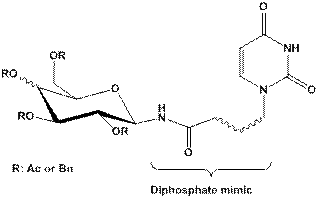
References:
[1] Lairson L.L., Henrissat B., Davies G.J., Withers S.G. (2008) Annu. Rev. Biochem. 77, 521–555.
[2] Berg S., Kaur D., Jackson M., Brennan P.J. (2007) Glycobiology 17, 35–56.
[3] Behr J.B., Gainvors-Claisse A., Belarbi A. (2007) Nat. Prod. Res. 21, 76–82.
Roman Komor received a scholarship under the project DoktoRIS - Scholarship Program for Innovative Silesia.
61. PREPARATION
OF ACYCLIC ANALOGUES OF URIDINE
AND THEIR APPLICATION IN SYNTHESIS OF POTENTIAL GLYCOSYLTRANSFERASE INHIBITORS
Roman Komor, Gabriela Pastuch, Wiesław Szeja
Silesian University of Technology, Department of Organic Chemistry, Bioorganic Chemistry and Biotechnology, 44-100 Gliwice, ul. Krzywoustego 4, Poland.
Oligosaccharides and glycoconjugates that are found on the cell surface participate in many intracellular and extracellular events such as viral or bacterial infection, tumor metastasis, immune response or inflammation. The development of glycosyltranferases inhibitors may lead to discovery of novel therapeutic agents for the treatment of certain diseases in which carbohydrate-protein interactions are involved. To find suitable and selective inhibitors for this class of enzymes is still challenging. Here, we describe a novel concept that allows a design of inhibitors based on the structure of the donor substrate binding pocket.
The aim of the research is to synthesize potential glycosyltransferase inhibitors, acyclic derivatives of uridine connected to thioglycosides via amide bond, that are analogues of natural donor substrate. Structures that we propose were carefully designed, concerning requirements for the acyclic part of the nucleoside: (1) chain no longer than six carbon atoms, (2) substituents containing heteroatoms with lone pair of electrons (hydroxyl groups are preffered), and (3) presence of the carboxyl group which is essential to create amide bond with the amino group of glycoside.
Synthesis of such models will complete the already existing structures library and subsequent biological evaluations will be enriched with information concerning the biochemical relevance of size and structure of ribose mimicking motif of the obtained glycoconjugates. Modulation or inhibition of glycosyltransferase activity provides an opportunity for intervention of the composition of carbohydrates in oligosaccharides which may clarify the structure-function relationship. Moreover compounds that can modulate the biosynthesis of glycoconjugates are of great interest as novel therapeutic agents.
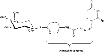
References:
[1] Zhu X., Stolz F., Schmidt R.R. (2004) J. Org. Chem. 69, 7367-7370.
[2] Vaghefi M.M., Bernacki R.J., Dalley N.K. et al. (1987) J. Med. Chem. 30, 1383-1391.
[3] Szewczyk B., Tyborowska J., Krol E., Szeja,W. (2005) J. Biotechnol. 118S1, S84-S85.
[4] Komor R., Pastuch-Gawołek G., Sobania A., Jadwiński M. (2012) Acta Polon. Pharmaceut. - Drug Res. 69, 1259-1269.
Roman Komor received a scholarship under the project DoktoRIS - Scholarship Program for Innovative Silesia.
62. Synthesis of dimeric uridine derivative – potential inhibitor of chitin synthase
Katarzyna Kral, Jadwiga Paszkowska, Tadeusz Bieg, Ilona Wandzik
Department of Organic Chemistry, Bioorganic Chemistry and Biotechnology, Silesian University of Technology, 44 –100 Gliwice, ul. Krzywoustego 4, Poland.
Chitin synthase (CS) is an enzyme responsible for fungal cell wall biosynthesis which is essential for fungal growth and reproduction. In the active site CS polymerizes uridine diphosphoryl-N-acetylglucosamine (UDP-GlcNAc) into polymeric chains of b-(1-4)-linked N-acetylglucosamine (GlcNAc) – chitin. The fact that chitin is absent in human cells makes CS an important antifungal target.
The structure of the active site in chitin synthase is still unknown, therefore potential inhibitors are designed based on the proposed mechanism. It is called “The two-active site mechanism” and it suggests the presence of two active sites in the enzyme with the distance about 14 – 22 Å between them. According to this hypothesis, CS simultaneously catalyses polymerization of chitin in these two places.
Several dimeric inhibitors of CS were synthesized and their biological activity showed advantage of dimeric compounds compared to monomer analogues.1
Following this theory we designed and synthesized dimeric inhibitor (Figure1). It contains two molecules of uridine with a proper linker between them. The synthetic procedure of this compound is described below.

Fig 1. Structure of synthesized dimeric derivative of uridine
The multistep synthesis of dimeric derivative utilizes 2’,3’-O-isopropylideneuridine and 5’-amino-2’,3’-di-O-tert-butyldimethylsilyluridine as main substrates. Both of them were first coupled with N-benzyloxycarbonylglycine, then protective groups were cleaved by hydrogenolysis to obtain derivatives with free amino group. In the next step reaction with succinic anhydride occurred and finally both fragments were coupled using PyBOP, which is commonly used as a coupling reagent2 in peptide bond formation. All compounds were purified by flash chromatography and characterized by NMR spectroscopy. Synthetic details will be presented.
References:
[1] Yeager A.R., Finney N.S. (2004) J. Org. Chem. 69, 613-618.
[2] Coste J., LeNguyen D., Castro B. (1990) Tetrahedron Lett. 31, 205-208.
Research studies part-financed by European Union within the European Regional Development Fund (POIG 01.01.02-14-102/09).
63. SYNTHESIS AND FUNGICIDAL ACTIVITY OF THIOCARBAMATES DERIVATIVES ALCOHOLS
Agata
Ptaszek-Budniok1, Anna Kasprzycka1, Przemysław Hahn1,
Wiesław Szeja1,
Wioletta Przystaś2, Ewa Zabłocka-Godlewska2, Mirosława
Sioła3
1Silesian University of Technology, Faculty of
Chemistry, Krzywoustego 4Str., 44-100 Gliwice, Poland; 2Silesian
University of Technology, Faculty
of Power And Environmental Engineering, Konarskiego 18 Str.,
44-100 Gliwice, Poland; 3Laboratory of Microbiology Silesian Center for Heart Diseases, M.
Curie-Skłodowskiej 9 Str., 41-800 Zabrze.
E-mail: agata.ptaszek-budniok@polsl.pl
N-Allyl and N-aryl tiocarbamates reported possess very interesting biological properties.1-3 Thiocarbamates used as fungicides have been extensively studied since 1960.4 However, little attention had been given to N-allyl and N-aryl substituted thionocarbamates derivatives alcohols until Thorne5 studied a wide variety of derivetives of thiocarbamic acid and carbamic acids. He found that phenyl carbamates, alkyl-phenyl carbamates, and alkyl thiophenyl carbamates have low fungitoxicity and that some thionocarbamates showed activity. To study the possibility of improving the fungicidal activity of the known thiocarbamates, in the present work a series of N-alkyl, N-phenyl and N-benzyl thiocarbamates derivatives alcohols were synthesized, and their fungitoxic activity was studied for test isolates: Phoma linganum, Fusarium solani, Fusarium culmorum, Trichoderma viride and Botrytis cinerea which were obtained from the Bank of Plant Pathogens (Institute of Plant Protection, National Research Institute in Poznań). In addition, thiocarbamates synthesized were tested for yeasts (Candida albicans, Candida dubliniensis, Candida tropicalis, Candida glabrata, Candida krusei, Candida parapsilosis) isolated from clinical materials at the Laboratory of Microbiology, Silesian Center for Heart Diseases in Zabrze.
Results presented in this paper reveal good fungicidal activity of certain N-allyl, N-phenyl and N-benzyl thiocarbamates.
References:
[1] Cesarini S., Spalarossa A., Ranise A., et al. (2008) Bioorg. Medic. Chem. 16, 6353-6363.
[2] Albores-Velasco M., Thorne J., Wain R.L. (1995) J. Agric. Food Chem. 43, 2260-2261.
[3] Hart B.W., Faiman M.D. (1995) Biochem. Pharmacol.,16,157-163.
[4] Ludwig R.A., Thorn, G.D. (1960) Adv.Pest Control Res. Interscience: New York, Vol.3.
[5] Thorne J. (1967) Chemical structure in relation to biological activity of certain carbamic acid derivatives. Ph.D Dissertation, University of London.
Research studies part-financed by the European Union within the European Regional Development Fund (POIG. 01.01.02-14-102/09).
64. SYNTHESIS OF NOVEL AND ACTIVE SUGAR-BASED DRUG DELIVERY SYSTEMS
Sylwia Waśkiewicz1, Małgorzata Burek1, Agata Grzybek1, Zenon P. Czuba2, Wojciech Król2
1Department of Physical Chemistry and Technology of
Polymers, Faculty of Chemistry, Silesian University
of Technology, ul. M. Strzody 9, 44-100 Gliwice, Poland; 2Department
of Microbiology and Immunology, Medical University of Silesia, 41-808 Zabrze,
ul. Jordana 19, Poland.
“Intelligent” hydrogels, which can change their swelling behavior and other properties in response to environmental stimuli such as temperature, pH, solvent composition and electric fields are interesting in view of their unique properties and potential application in the technological and biomedical fields. Temperature and pH are two important parameters that are normally studied in detail for drug delivery purposes. It is necessary for such systems to show a response at body temperature and under acidic pH in order to function as desired at the target area. Among the family of temperature-sensitive hydrogels, poly(N-isopropylacrylamide) (PNIPAAm) hydrogel is one of the most widely studied. The hydrogels based on PNIPAAm exhibit the volume phase transition temperature (VPTT) around 32°C, which means that above this temperature their nature changes from swollen to skrinked. In other words, hydrogels show an on-off drug release, allowing the desired drug release above VPTT. However, their disadvantages are that they mostly show a slow response to external stimuli and are usually non-degradable, which may restrict their applications as biomaterials. One way to improve the response rate and simultaneously biocompability of PNIPAAm based hydrogels is through the introduction of sugar moieties. Moreover, sugar units can modify VPTT of PNIPAAm hydrogel to the body temperature.
In the present study we report the synthesis of a series of novel sugar based thermoresponsive hydrogels based on trehalose or salicin. Those hydrogels are proposed to be used as a drug delivery carriers, releasing drug above VPTT with predetermined pharmacokinetics. In the first case acid-degradable trehalose-based diacetals were used as a cross-linker. Hydrogels structure is destroyed in an acidic environment as a result of deprotection reaction and is accompanied by a release of trehalose molecules. Trehalose is a storage carbohydrate and transport sugar and plays an important role in stress protection, especially during heat stress and dehydratation. As a non-reducing sugar, this saccharide does not undergo Maillard reaction with amino compounds such as amino acids or proteins. It is used in protecting corneal epithelial cells in culture from death by desiccation and suppression of tissue denaturalization, for effective preservation of organs, in the treatment of osteroporosis and in protein-based drugs including Advate (Baxter), Avastin (Genentech), Lucentis (Genentech), and Herceptin (Genentech).
Similar hydrogels were obtained using salicin-monoacetal compounds, where a salicin molecule is released in an acidic environment without hydrogel structure degradation. Salicin, is an active precursor for salicic acids, used as anti-inflammatory drug and could be employed instead of aspirin.
The preliminary cytotoxicity studies by employing MTT assay have shown that sugar acetals, their hydrogels and copolymers with NIPAAm are not cytotoxic for the human fibroblasts cell line.
The work is financed by the POMOST Programme 2012-5/11 of the Foundation for Polish Science and co-financed by the European Regional Development Fund.
Cellular Stress, Oxidation
and DNA Repair
65. THE PARTICIPATION OF WIP1 PHOSPHATASE IN THE RESPONSE
OF U-2 OSTEOSARCOMA CELLS TO RADIATION
Agata
Abramowicz1, Katarzyna Szołtysek1, Patryk Janus1,
Krzysztof Puszyński2,
Piotr Widłak1
1Maria Skłodowska-Curie Memorial Cancer Center and Institute of Oncology, Gliwice, Poland; 2Silesian University of Technology, Gliwice, Poland.
WIP1 is a multi-target serine/threonine protein phosphatase encoded by PPM1D gene. Its major targets include MDM2 (Ser395), ATM (Ser1981), p53 (Ser15), CHK1 (Ser345), CHK2 (Thr68), γH2AX (Ser139) and NF-κB (Ser536). These proteins are inactivated by a WIP1-mediated dephosphorylation, hence WIP1 putatively participates in regulation of recovery of cell after induction of DNA damage response (DDR). Overexpression of PPM1D gene has been observed in some types of cancer and usually has been associated with poorer prognosis. Here, we aimed to characterize the significance of WIP1 in response to radiation in human osteosarcoma U-2 OS cell line.
U-2 OS cells have been characterized by high expression of PPM1D gene. Two functional gene products are detected in these cells: full-length wild-type protein (66.7 kDa) and C-terminus truncated form (about 50 kDa). The expression of WIP1 in U-2 OS cells was inhibited by lentivirus-delivered shRNA (control cells contained an shRNA construct encoding a scrambled sequence). The WIP1 knock-down was verified by Western-blot – amounts of both protein forms were reduced to approximately 25% of their initial levels. The DDR was induced upon exposure to ionizing radiation (IR, 4 or 10 Gy) or UV-C radiation (10 J/m2). The levels of DDR-related proteins were analyzed by Western-blot at different time points after irradiation. Expression of selected p53-dependent genes (NOXA, CDKN1A) was assessed by QRT-PCR. The influence of the WIP1 inhibition on the cell cycle profiles was assessed by flow cytometry. The long-term effects of the WIP1 deficiency on radiation sensitivity of cells were analyzed by the clonogenic test.
Both IR and UV-C induced accumulation of WIP1 in control cells, which was the most prominent after irradiation with 10 Gy, also in WIP1-depleted cells some accumulation of WIP1 could be detected after exposure to IR.
The WIP1 deficiency in U-2 OS cells resulted in prolonged phosphorylation of Chk1 (Ser345) and Chk2 (Thr68) in UV and IR exposed cells, respectively. The effect was visible 24 hours after irradiation, which corresponded to maximum accumulation of WIP1 in WIP1-proficient cells. It suggests that WIP1 could be involved in long-term “switch-off” of cellular response to radiation. Furthermore, late activation/phosphorylation of Chk2 (Thr68) could be detected in UV-irradiated WIP1-deficient cells, hence WIP1 could be involved in regulation of late/secondary response to UV-induced damage. However, similar distribution of the cell cycle and expression of p53-dependent genes in WIP1-proficient and -deficient cells upon irradiation indicates existence of mechanisms capable of general compensation of WIP1 deficiency.
This work was supported by Polish National Science Center, Grant N N518 287540.
66. CHARACTERIZATION OF ERCC1 DEFICIENT MOUSE EMBRYONIC FIBROBLASTS IN
RESPONSE TO 4-HYDROXY-2-NONENAL, A MAJOR
LIPID PEROXIDATION PRODUCT
Jolanta Czerwińska1, Patrycja Berentowicz2,
Małgorzata Nowak2, Konrad Kosicki2,
Laura Niedernhofer3, Elżbieta Speina1, Barbara Tudek1,2
1Institute of Biochemistry and Biophysics, Polish Academy of Sciences, Warsaw, Poland; 2Institute of Genetics and Biotechnology, Warsaw University, Warsaw, Poland; 3The Scripps Research Institute - Florida, Jupiter, USA.
The ERCC1-XPF protein complex is an endonuclease involved in nucleotide excision repair (NER) and processing of DNA interstrand crosslinks. Ercc1 knockout mice display retarded postnatal growth and premature aging. Cells derived from Ercc1-/- specimens reveal an increased level of reactive oxygen species and are hypersensitive to factors introducing oxidative stress. It was shown that during oxidative stress secondary products of reactive oxygen species – lipid peroxidation (LPO) products are intensively generated. The aim of our study was to determine the role of LPO products in etiology of the Ercc1-/- mice phenotype. For this purpose, immortalized mouse embryonic fibroblasts (MEFs) derived from Ercc1-/- and wild type (wt) mice were used. We investigated the sensitivity of Ercc1-/- and wt MEFs to hydrogen peroxide (H2O2) and four aldehydes – end products of LPO: 4-hydroxy-2-nonenal (HNE), crotonaldehyde (CRO), malondialdehyde (MDA) and acrolein (ACR). These compounds are very reactive and can form adducts to DNA and proteins as well as DNA-DNA and DNA-protein crosslinks. Our results demonstrated hypersensitivity of Ercc1-/- fibroblasts to H2O2, as well as to LPO products: HNE, CRO and MDA, but not to ACR. The biggest difference in viability between Ercc1-/- and wt cells was in the case of HNE treatment. Since HNE is also one of the major LPO products generated in cells, we chose this compound for further study aimed at characterization of cell death induced in Ercc1-/- MEFs by LPO products. Three types of cell death: apoptosis, necrosis and autophagy were investigated by luminescence assays and/or by Western blotting. Necrosis and apoptosis were the preferred types of HNE-induced cell death for Ercc1-/- and wt, respectively. There was no activation of autophagy. We also measured the cell cycle of Ercc1-/- MEFs by flow cytometry and found in response to HNE a marked accumulation of fibroblasts in phase G2.
67. THE COMET ASSAY AS BIOMARKER OF DNA DAMAGE
IN PERIPHERAL BLOOD LEUKOCYTES OF NUCLEAR MEDICINE STAFF
Małgorzata M. Dobrzyńska1, Krzysztof A. Pachocki1, Aneta Gajowik1 Joanna Radzikowska1, Agata Sackiewicz-Słaby2
1Department of Radiation Protection and Radiobiology,
National Institute of Public Health – National Institute of Hygiene, 00-791
Warsaw, Chocimska 24, Poland; 2Maria Sklodowska Curie Memorial
Center and Institute
of Oncology ,02-781 Warsaw, Roentgena 5, Poland.
High, but also low chronic doses of ionizing radiation are known to be mutagenic and carcinogenic in humans. Medical staff using radiation for diagnostic and therapeutic purposes are potentially at risk of overexposure.
The purpose of the study was the examination of DNA damage in peripheral blood leukocytes of staff at the Department of Nuclear Medicine of two medical centers in Warsaw, where several kinds of radionuclides are used for radiodiagnosis and radiotherapy. Control were chosen among unexposed staff. Both exposed and control individuals were asked to fill up a questionnaire to get necessary information about sex, age, smoking habits, exact description of work, use of therapeutic drugs, previous diagnostic exposure to X-rays, nuclear medicine examination. The blood was taken by venipuncture. We used the alkaline Comet assay to assess the DNA damage. Percentage of DNA in the tail and Comet tail moment were used for further analysis.
The results show variability between individuals from both control and exposed groups. The significantly higher mean of DNA damage in lymphocytes of investigated group compared to unexposed control was observed. Among exposed staff there were differences dependent on the kind of work. Mean percentage of DNA in Comet tail and Comet tail moment of technicians involved in scintigraphy and positron emission tomography markedly exceeded the mean values of other group. The level of DNA damages of lymphocytes of nurses, doctors, pharmacists and administrative workers (contact only with treated patients) was much lower. The level of DNA damage of peripheral blood leukocytes was independent on the period and category of work of exposed people. Cigarette smoking was not related to increases in DNA damage in white blood cells of nuclear medicine personnel in contrary to DNA damage of control individuals. There were no differences between females and males among control and exposed subjects.
In conclusion, monitoring and improvement of health safety of nuclear medicine personnel should be taken under consideration to prevent induction of DNA strand breaks.
This work was funded by Ministry of Science and Higher
Education/National Centre for Research
and Development, Project “Improvement of Safety and Working Conditions”
coordinated by Central Institute for Labour Protection-National Research
Institute (2011-2013).
68. 8-Oxo-7,8-dihydroguanine (8oxoGua) and uric
acid
as an efficient predictors of survival in colon cancer (CRC) patients.
Relationship between the main enzymes (PARP and OGG) responsible for 8oxoGua
repair in CRC
Tomasz Dziaman1, Marek Foksiński1,
Daniel Gackowski1, Rafał Różalski1,
Ewelina Zarakowska1, Anna Szpila1, Jolanta Guz1,
Beata Sikorska1, Agnieszka Siomek1, Hubert
Ludwiczak2, Jarosław M. Cieśla2, Mateusz Chmielarczyk2,
Ewa Wiśniewska3, Andrzej Marszałek3,4, Barbara Tudek2,5,
Ryszard Oliński1
1Department of Clinical Biochemistry, Collegium
Medicum, Nicolaus Copernicus University, Karlowicza 24, Bydgoszcz, Poland; 2Institute of Biochemistry and Biophysics, Polish
Academy of Sciences, Pawinskiego 5a, Warsaw, Poland; 3Department of Clinical Pathomorphology, Collegium
Medicum, Nicolaus Copernicus University, Sklodowskiej-Curie 9, Bydgoszcz,
Poland; 4Department of Oncologic Pathology, University
of Medical Sciences and Wielkopolskie Oncology Center, Garbary 15, 61-868
Poznan, Poland; 5Institute
of Genetics and Biotechnology, Faculty of Biology, University of Warsaw,
Pawinskiego 5a, Warsaw, Poland.
The aim of this work was to answer the question whether the broad range of parameters which describe oxidative stress and oxidatively damaged DNA and repair are appropriate prognosis factors of survival for colon cancer patients (CRC)? The following parameters were analyzed for 89 CRC patients: concentration of uric acid and vitamins A, E, C in plasma; levels of 8-oxodG in DNA of leukocytes and colon tissues; urinary excretion rates of 8-oxodG and 8-oxoGua; the activity and mRNA or protein level of repair enzymes OGG1, APE1, ANPG, TDG and PARP1.
All DNA modifications and plasma antioxidants were analyzed using high performance liquid chromatography (HPLC) or HPLC/gas chromatography-mass spectrometry techniques. Expression of repair proteins was analyzed by QPCR, Western or immunohistochemistry methods.
Longer survival coincided with low levels of 8-oxodG/8oxoGua in urine and 8-oxodG in DNA as well as with high concentration of uric acid plasma level. In contrast to expectations, longer survival coincided with lower mRNA level in normal colon tissue of the main 8-oxoGua DNA glycosylase, OGG1, but no association was found for PARP-1 expression.
When analyzing two parameters simultaneously, the discriminating power increased significantly. Survival prognosis for patients with low level of urinary 8-oxoGua together with low level of 8-oxodG in leukocytes (both below median value) or high concentration of plasma uric acid (above median value) have the best prediction power. Since prediction value of these parameters seems to be comparable to conventional staging procedure, they could possibly be used as markers to predict clinical success in CRC treatment.
In our work we have also observed statistically highly significant correlation between mRNA expression of OGG1and PARP-1 in all investigated tissues/settings; leukocytes of all groups (control, patients with adenoma and CRC) and in marginal/healthy tissues as well as in cancerous one. The good correlation may indicate that both genes, which are main players in repair of oxidatively damaged DNA, are commonly expressed as an answer to the same stimuli – oxidative stress, which is a feature of CRC.
Interestingly, we observed significant correlation between protein expression of PARP-1 and OGG1only in diseased tissues – polyps and cancerous one. This in turn may be explained by the finding that direct interaction of the proteins is significantly enhanced by oxidative stress.
69. ANTICANCER PROPERTIES OF FERROCENYL DERIVATIVES:
DNA DAMAGE IN
HUMAN HEPATOMA CANCER CELLS HEPG2 TREATED
WITH ACRYLOYLO FERROCENES
Paweł
Hikisz1, Paulina Lewarska1, Łukasz Szczupak2,
Konrad Kowalski2,
Aneta Koceva-Chyła1
1Department of Thermobiology, Institute of Biophysics,
Faculty of Biology and Environmental Protection, University of Lodz, Pomorska
141/143, 90‐236 Lodz, Poland; 2Department of Organic
Chemistry, Institute
of Chemistry, Faculty of Chemistry, University of Lodz, Tamka 12, 91-403 Lodz,
Poland.
Metal complexes with anticancer properties make up a part of new trends in contemporary oncology. High hopes for the treatment of cancer are linked with ferrocene derivatives. Their chemical stability, hydrophobicity and lipophilicity, easy chemical modification and functionalization, as well as strong redox properties make them suitable candidates as potential chemotherapeutic agents. Cytotoxic properties of ferrocene correspond to their ability to generate free radicals and DNA cleavage activity thus induce oxidative damage to DNA and leads to strand breaks in deoxyribonucleic acid.
The aim of this study was to investigate the effect of 3 ferrocene derivatives (15, 15Cl and 12a) on the generation of DNA damage in HepG2 human liver cancer cells. The single cell electrophoresis/comet assay, performed under alkaline conditions, was used to measure DNA damage. The cells were incubated for 24 hours with IC50 concentration of ferrocenes cells and after then cultured in fresh medium for 0 and 24 hours.
The results indicated that investigated ferrocenes derivatives cause DNA damage in HepG2 cells. Ferrocenes containing chlorine atom in their structure showed stronger genotoxic properties. After 24 hours of cell culture in drug free medium, a decrease in the amount of damaged DNA was observed compared with 0 h timepoint. This may suggest that in this cancer cell line at lest a part of DNA damage caused by investigated ferrocene derivatives are easily repaired by efficient DNA repair systems within a short time after the treatment.
70. MAJOR LIPID PEROXIDATION PRODUCT 4-HYDROKSYNONENAL AFFECTS DNA DAMAGE RESPONSE IN HeLa CELLS
Konrad
Kosicki1, Jolanta Czerwińska2, Jakub Kucharczyk1,
Elżbieta Speina2,
Wojciech Niedźwiedź3, Barbara Tudek1,2
1Institute of Genetics and Biotechnology, University of Warsaw, Pawińskiego 5a, Poland.;
2Institute of Biochemistry and Biophysics,
Polish Academy of Sciences, Pawińskiego 5a, 02-106 Warsaw, Poland; 3The Weatherall Institute of Molecular
Medicine, University of Oxford, John Radcliffe Hospital, Headington, Oxford OX3
9DS, UK.
Lipid peroxidation (LPO) is involved in the pathogenesis of several human diseases, including cancer. LPO products react with cellular proteins changing their properties, and with DNA bases to form mutagenic exocyclic-DNA adducts, DNA-DNA and DNA-protein crosslinks. We have shown that HNE induced single- (SSB) and double-strand DNA breaks (DSB) in HeLa cells, which number increased with time up to 24 h after cells treatment with HNE. SSB and DSB caused replication stress should induce the Fanconi system. However, FANCD2 ubiquitination was not induced following HNE treatment. Activation of Fanconi pathway is performed by phosphorylation of FANC core proteins by ATM/ATR kinases. We observed that HNE inhibits phosphorylation of several DNA Damage Response (DDR) proteins, namely Chk1, SMC1, KAP1, RPA, and histone H2AX. This inhibition was visible up to 12 h following cells’ exposition to HNE. However, phosphorylation of Chk1 was inhibited for the whole time of conducting the experiment, that is 72 h. Only very faint bands of P-Chk1 were seen 24-72 h after HNE treatment of HeLa cells. This could suggest that ATR kinase phosphorylating Chk1 is inhibited by HNE. Since Chk1 is the only kinase, which phosphorylates one of FANC core proteins FANCE, and this phosphorylation is indispensable for FANC core activation and FANCD2 ubiquitination, Chk1 inhibition could explain lack of FANC system activation following HNE treatment in spite of the presence of DNA damage. Interestingly, when HeLa cells were pre-treated with HNE, DDR was inhibited, when cells were subsequently exposed to another DNA damaging agent, camptothecin. DDR inhibition by HNE resulted in cell cycle arrest in G2 phase, and increase of DNA fragmentation. However, 6 h following HNE treatment phosphorylation of DNA-PKcs was induced. This could suggest that in response to HNE high fidelity homologous recombination is substituted by low fidelity non-homologous end joining, thus decreasing genome integrity. Thus, genotoxic activity of HNE is complex and involves both DNA damage and modulation of DNA damage response.
71. Quercetin triggers reactive oxygen
species-dependent induction of apoptosis and lysosomal death of sensitive
and multidrug resistant leukaemia HL60 cells
Agnieszka Maruszewska , Jolanta Tarasiuk
Department of Biochemistry, Faculty of Biology, University of Szczecin, 3c Felczaka St, 71-412 Szczecin, Poland.
Multidrug resistance (MDR) is one of the major causes of the failure of anticancer therapy. One of the most important mechanisms leading to the occurrence of MDR is related to the modulation of cellular death pathways. Reactive oxygen species (ROS) are among the most crucial factors triggering programmed cell death by apoptosis or lysosome membrane permeabilization-dependent mechanisms.
Polyphenols are compounds widely distributed in common diet (e.g. in fruits, vegetables, nuts, wine and tea). Quercetin belongs to the flavonoids – one of the subclasses of polyphenols and there is an increasing body of evidence showing that it is able to modulate cellular death pathways of tumour cells.
The aim of this study was to determine the effect of quercetin on triggering the programmed death of human promyelocytic leukaemia sensitive cells HL60 and multidrug resistant HL60/VINC cells overexpressing P-glycoprotein.
It was found that quercetin exerts comparable cytotoxic activity towards sensitive HL60 and resistant HL60/VINC cells. It has modulated the cellular level of ROS acting as prooxidant or antioxidant agent depending on the concentration used and time of incubation. The results of cell cycle distribution analysis showed significant increase (to about 40-50 %) in the percentage of sub-G1 subpopulation of cells exposed to quercetin used at IC90 in the case of both HL60 and HL60/VINC cells. It was also demonstrated that this compound caused oligonucleosomal DNA fragmentation characteristic for apoptosis. It caused significant activation of caspase-3 (1.5-fold in sensitive HL60 and 3.6-fold in resistant HL60/VINC cells) as well as caspase-8 (2.6-fold in sensitive HL60 and 5.6-fold in resistant HL60/VINC cells). Quercetin induced also lysosome membrane permeability of both HL60 and HL60/VINC cells in a concentration-dependent and time-dependent manner.
In summary, the obtained results show that quercetin is able to trigger ROS-dependent induction of apoptosis and lysosomal death of sensitive and multidrug resistant leukaemia HL60 cells.
This study was supported by the Faculty of Biology, University of Szczecin, Poland (Grant no. 504-1000-240-850).
72. Effect of flavonoid glycosides on reduction of superoxide level and DNa damages after X-ray radiation of human lymphocytes in vitro
Małgorzata Materska1, Irena Perucka1,
Maria Konopacka2, Jacek Rogoliński2,
Krzysztof Ślosarek3, Barbara Chilczuk1
1Research Group of Phytochemistry, Department of
Chemistry, Agricultural University, Akademicka 15,
20-950 Lublin, Poland; 2Center of Translational Research and
Molecular Biology of Cancer; 3Radiotherapy
and Brachytherapy Treatment Planning, Institute of Oncology, ul. Wybrzeże Armii
Krajowej 15, 44-100 Gliwice, Poland.
The antioxidant and radioprotective effects of four main glycosides of phenolic compounds of Capsicum annuum L. were studied. Sinapoyl-E-glucoside; quercetin-3-O-rhamnoside-7-O-glucoside, quercetin-3-O-rhamnoside and luteolin-7-O-(2-apiosyl)-glucoside were isolated and purified by preparative liquid chromatography and their structure was confirmed by spectroscopic methods (UV, ESI-MS). The compounds were assayed for the first time for their radioprotective effect from oxidative damage induced by X radiation using human lymphocytes. Simultaneously, antioxidant activity, with respect to superoxide radical generated by enzymatic and nonenzymatic methods, was determined. Glycosides of quercetin, luteolin and sinapic acid showed higher radioprotective activity than their aglycones, even though they showed weaker antiradical activity. The highest radioprotective potential was noticed for quercetin-3-O-rhamnoside. Furthermore, quercetin and luteolin derivatives, in contrast to the free aglycones, were not cytotoxic against human lymphocytes in the whole range of concentrations used. The obtained results indicate that derivatives of flavonoid are safer and more efficient in protecting cellular components from harmful effects of X-rays in comparison to their aglycones. These compounds can potentially be used as cell protective substances.
73. MODIFICATION OF WRN PROTEIN WITH 4-HYDROXYNONENAL
CHANGES ITS ENZYMATIC ACTIVITIES AND FUNCTIONAL DYNAMICS
Elżbieta Speina1, Jolanta
Czerwińska1, Jarosław Poznański1, Janusz Dębski1,
Vilhelm A. Bohr2, Barbara Tudek1,3
1Department of Molecular Biology, Institute of
Biochemistry and Biophysics, Polish Academy of Sciences,
02-106 Warsaw, ul. Pawińskiego 5a, Poland; 2Laboratory of Molecular Gerontology,
National Institute
on Aging, National Institutes of Health, 21224 MD, Baltimore, 251 Bayview Blvd, USA; 3Institute of Genetics and
Biotechnology, Warsaw University, 02-106 Warsaw, ul. Pawińskiego 5a, Poland.
4-Hydroxy-2-nonenal (HNE) is a reactive a α,β-unsaturated aldehyde produced during oxidative stress and subsequent lipid peroxidation of polyunsaturated fatty acids. The reactivity of 4-HNE towards DNA has been well established. Here, Werner (WRN) protein is identified as a target for modification by 4-HNE. Werner syndrome arises through mutations in both copies of the WRN gene that encode RecQ 3’-5’ DNA helicase and exonuclease essential for genomic stability. This hereditary disease is associated with chromosomal instability, premature aging and cancer predisposition. WRN appears to participate in the cellular response to oxidative stress and cells devoid of WRN display elevated levels of oxidative DNA damage.
Western blot, immunoprecipitation and mass spectrometry were used to identify and characterize the in vitro and in vivo covalent modifications of WRN by HNE. Sites of 4-HNE adduction on human recombinant WRN were mapped as Lys577 (within ATP binding domain), Cys727, His1290, Cys1367, Lys1371 (within nuclear localization signal) and Lys1389. HNE adduction of WRN protein was shown to appear also in vivo, in cells pretreated with HNE or carbon tertrachloride (the agent known to cause lipid peroxidation), suggesting that this modification could influence WRN catalytic activities. We demonstrated that both helicase and exonuclease activities of HNE modified WRN protein were inhibited both in vitro and in immunoprecipitates from cell extracts. However, WRN binding to DNA was unaffected. We applied molecular modeling analysis of HNE adducted to Lys577 and Cys727 residues to provide a potential mechanism of deregulation of WRN enzymatic activity.
74. Involvement of Reactive oxygen species and microRnas
in cellular response to oxidative stress
Izabella Ślęzak-Prochazka1,2, Karolina Gajda1, Roman Jaksik1, Joanna Rzeszowska-Wolny1
1Institute of Automatic Control, Silesian University of Technology, 44- 100 Gliwice, Akademicka 16, Poland; 2Department of Public Health, Czestochowa University of Technology, 42-200 Częstochowa, Armii Krajowej 36b, Poland.
MicroRNAs (miRNAs) are small (~22nt) noncoding RNA molecules that negatively regulate gene expression by binding to the 3’untranslated region (3’UTR) of their target mRNAs. MiRNAs play an important role in cellular processes like apoptosis, proliferation and stress response. It is evident that miRNAs are involved in regulation of the biological pathways associated with ionizing radiation-induced stress responses. However, the exact mechanisms causing altered miRNA levels and the direct targets of many stress-induced miRNAs are largely unknown. The aim of this project is to determine miRNA contribution to cellular response to ionizing radiation.
Leukemic K562 and HL60 and melanoma Me45 cell lines were exposed to ionizing radiation. We determined levels of reactive oxygen species (ROS) using 2′,7′-dichlorofluorescein diacetate (DCFH) and analyzed cell cycle distribution using propidium iodide 1, 4, 8, 12 and 24h after irradiation. In both K562 and HL60 cells, the highest increase in ROS levels was observed 12h after irradiation compared to control non-irradiated cells, whereas we did not observe this increase in Me45 cells. K562 and HL60, but not Me45 cells showed cell cycle arrest in G2/M phase 12h after irradiation. Next, to investigate whether ROS increase is dependent on cell cycle phase, we arrested K562 cells by nocodazole or thymidine block and we observed a similar G2/M-specific increase of ROS detected by DCFH. To determine the involvement of miRNAs, we compared number of predicted miRNA target genes in transcripts up- or downregulated 1, 12 and 24h after irradiation. We observed changes in the number of miRNA target genes 12h after irradiation in K562 cells, but not in Me45 cells.
These results suggest that miRNAs may cooperate with ROS to induce cellular response to ionizing radiation and regulate cell cycle.
This work was financially supported by Grant No. DEC-2012/04/A/ST7/00353, National Science Centre (NCN) and BKM-514/Rau1/2013/t26, Silesian University of Technology.
Normal Cell Biology
75. PROINFLAMMATORY CONDITIONS STIMULATE CD274 EXPRESSION
ON THE HSCS
Agnieszka Ciomber1,2, Iwona Mitrus1, Magdalena Głowala-Kosińska1, Wojciech Fidyk1, Andrzej Smagur1, Sebastian Giebel1
1Department
of Bone Marrow Transplantation, Maria Skłodowska-Curie Memorial Cancer Center
and Institute of Oncology, Gliwice Branch, 44-101 Gliwice, Wybrzeże Armii
Krajowej 15, Poland; 2Department of Animal Physiology and
Ecotoxicology, Faculty of Biology and Environmental Protection, University of
Silesia,
40- 007 Katowice, ul. Bankowa 9, Poland.
Hematopoetic stem cells (HSCs) are responsible for the production of all the lineages of blood cells. HSCs reside inside the bone marrow microevironment, where several groups of cells and extracellular matrix elements interact with HSCs to regulate their self renewal, differentiation and migration. HSCs may directly interact with the immune system. For instance, they possess the ability to regulate expression of CD274 proteins, which is believed to have immunosupressive function. Cell‑surface CD274 inhibits T-cell immunity by induction of apoptosis in T lymphocytes. Expression of this protein on HSCs can be induced by stress or immune signals but this process is still poorly described.
The aim of the study was to analyze the expression of CD274 on the surface of HSCs. Firstly, we compared the percentage of CD274+ HSCs in bone marrow samples obtained from patients before allogeneic HSCs transplantation and 28 days after the procedure. Besides, in the in vitro culture we investigated the impact of proinflammatory and immmunosupressive cytokines on the level of CD274 on the HSCs obtained from peripheral blood stem cells (PBSCs). We examined the effect of IL-2, IL‑4, IL-10, TGF-β and IFN-γ after 24, 48 and 72 hours incubation. The analysis were performed using flow cytometry and appropriate antibodies.
Our results demonstrate increased level of CD274 protein on HSCs after HSCs allotransplantation. The percent of CD274 positive HSCs is 2- fold higher than the one before transplantation. In vitro studies show that proinflammatory cytokines stimulate CD274 expression on HSCs. The percent of CD274+ HSCs increases after 24 hours of incubation from 3 to 25% under the influence of IL-2 and to 75% after stimulation with IFN-γ the effect maintains or increases over time. There are no changes in the level of this protein after stimulation with IL-4, IL-10 and TGF-β which have immunosuppressive properties. We assume that the increased expression of CD274 on HSCs is associated with inflammatory conditions in bone marrow microenvironment after HSCs transplantation. The higher expression of this protein probably allows them to survive in recipient’s microenvironment.
The study was supported by Grant No. 2011/03/B/NZ6/04917 KTS.
76. Influence
of TX-100, SDS and trypsin on viability
of fibroblasts in vitro
Michalina Gramatyka, Piotr Wilczek
Foundation of Cardiac Surgery Development, 41-800 Zabrze, Wolności 345a.
Heart valve diseases are an important mortality cause in patients suffering from cardiovascular diseases. The mechanical and biological valve prostheses, which are currently used, show many limitations associated with their fast degradation (in case of biological prostheses) or ability to cause thromboembolic complications (in case of mechanical prostheses). Removal of the donor cells (decellularization) from tissue and replacing them with recipient cells is the promising method to overcome these limitations. Such approach presents a number of potential advantages including: reduction of the immune response, reduction of the valve calcification intensity and reduction of the risk of transmitting diseases.
Most common methods of tissue decellularization include detergent and/or enzymatic treatment. Because all compounds used in the decellularization process show some degree of cytotoxicity, the question arises if their remains in the tissue can negatively affect condition of reseeded cells.
Here we aimed to assess toxicity of trypsin, SDS and Triton X-100 at concentrations corresponding to that used for decuellularization of heart valaves (for trypsin 1, 2 and 3 µg/ml; for SDS 10, 20 and 30 µg/ml; for Triton X-100 2, 4 and 6 µg/ml). We tested influence of these reagents on viability (TUNEL, JC-1 and AnnexinV tests), proliferation (Ki-67 test), adhesion (vinculin, ICAM-1, PECAM-1, VCAM-1 and E-cadherin proteins) and morphology (actin, β-tubulin and vimentin proteins) of fibroblasts cultured in vitro, .
We observed that for all concentrations and incubation times neither trypsin nor SDS nor Triton X-100 affected significantly viability of fibroblasts. Similarly, no effect on expression of cytoskeleton and adhesion proteins was observed. There was a slight decrease in proliferation capacity when incubating the cells with the highest concentration of Triton X-100. Further studies with different cell types could allow to confirm (or deny) if compounds remaining in the tissue after the decellularization process may have adverse effect on the condition of the recipient cells.
77. AN INTRIGUING ROLE OF  HIF1α  IN REGULATION OF

IN KERATINOCYTES
Anna Habryka1,2, Agnieszka Gogler-Pigłowska1, Mariusz Kryj3, Zdzisław Krawczyk1,2, Katarzyna Klarzyńska1,4, Dorota Ścieglińska1
1Center for Translational Research and Molecular
Biology of Cancer, 3Oncologic Surgery Clinic, Maria Skłodowska-Curie
Memorial Cancer Center and Institute of Oncology, Gliwice Branch, Poland; 2Department
of Organic Chemistry, Bioorganic Chemistry and Biotechnology, Faculty of
Chemistry, Silesian University
of Technology, Poland; 4Department of Biochemistry and Medical
Genetics, School of
Health Sciences, Medical University of Silesia, Katowice, Poland.
Epidermis, the most superficial layer of the skin, is composed mostly of keratinocytes. It is widely known that keratinocytes environment is hypoxic due to a lack of vasculature system in the epidermis. This phenomenon is manifested by stabilization of HIF1α in the basal layer, the most hypoxic part of the epidermis. Interestingly, as demonstrated by our previous results, HSPA2 is highly expressed in the epidermal basal layer.
We have performed an in silico analysis of the potential transcription factors binding sites in the HSPA2 gene promoter identifying the presence of Hypoxia Responsive Element (HRE), HIF1α binding regulatory sequence, as a result. Consequently, our goal was to investigate the potential role of HIF1α in regulation of the HSPA2 gene expression in human keratinocytes.
We have determined that during hypoxia in normal human epidermal keratinocytes (NHEK), as well as in immortalized human keratinocytes HaCaT (human adult low calcium high temperature) and in squamous cell carcinoma (A431) cell expression of the HSPA2 gene is downregulated. Functional analysis of HSPA2 promoter confirmed these results, showing decreased promoter activity under hypoxic conditions. Furthermore, deletion of HRE from the HSPA2 promoter significantly reduced activity of reporter Luc gene both under hypoxia and normoxia. This result suggests that HRE is required to maintain the expression of HSPA2 gene at normoxia. In Chromatin Immunoprecipitation assay (CHIP) we confirmed that HIF1α binds to HSPA2 promoter in keratinocytes cultured at low oxygen tension. In order to confirm whether HIF1 binding reduces HSPA2 promoter activity at hypoxia, we are currently performing chemical inhibition of HIF1α activity using N-acetylocystein (NAC), which reduces HIF1α stability and echinomycin, which specifically blocks interaction of HIF1α with DNA.
Our results enabled us to draw an overall conclusion that HIF1α acts as a negative regulator of the HSPA2 promoter activity in keratinocytes under hypoxia.
Research was financed from MNiSW grant No. N401 683740 and NCN grant No. N/NZ1/05/2012/00022. 78. HSPA2 IS A MARKER OF EPIDERMAL UNDIFFERENTIATED KERATINOCYTES
Katarzyna Klarzyńska1,3, Anna Habryka1,2, Agnieszka Gogler-Pigłowska1, Mariusz Kryj3, Zdzisław Krawczyk1,4, Damian Sojka1, Dorota Ścieglińska1
1Center for Translational Research and Molecular
Biology of Cancer; 3Oncologic Surgery Clinic, Maria Skłodowska-Curie
Memorial Cancer Center and Institute of Oncology, Gliwice Branch, Poland; 2Department
of Organic Chemistry, Bioorganic Chemistry and Biotechnology, Faculty of
Chemistry, Silesian University
of Technology, Poland; 4Department of Biochemistry and Medical
Genetics, School of
Health Sciences, Medical University of Silesia, Katowice, Poland.
This study was designed to characterize the expression of HSPA2 in human epidermis and during process of keratinocytes differentiation. HSPA2 was originally described as testis-specific member of the HSPA (HSP70) heat shock protein family, which is crucial for spermatogenesis. However, recently we have shown that HSPA2 protein is synthesized in human normal somatic tissues in cell- and tissue-type specific manner. High level of HSPA2 protein was found in basal layer of epidermis and other stratified epithelia. Nevertheless, possible function of HSPA2 and mechanisms regulating its expression in epidermis remain to be elucidated.
In the present study, by means of immunofluorescence analysis, we have demonstrated that in human epidermis HSPA2 positive cells colocalize with cells expressing markers specific for undifferentiated keratinocytes (CK5 and CK14). Therefore, we assumed that HSPA2 expression can be regulated during keratinocytes’ differentiation. We applied the model of calcium-induced differentiation of HaCaT cells to search for relation between the level of HSPA2 protein and keratinocytes differentiation. HaCaT cells grown in low calcium medium (LCM) showed marker signature specific for undifferentiated (basal) keratinocytes, reduced proliferation rate and significant upregulation of HSPA2 expression. On the contrary, HaCaT cells cultured in high-calcium medium (HCM) showed upregulation of differentiation markers, elevated proliferation rate and stable HSPA2 transcript level. Our results also suggested that the final level of HSPA2 protein can be determined by so far unidentified posttranslational mechanism. We have observed accumulation of HSPA2 protein in high density cell culture which, surprisingly, was not accompanied by increase in the level of transcription.
In summary, our results strongly suggest that HSPA2 gene is highly active in undifferentiated keratinocytes (possibly epidermal progenitor and/or stem cells). In order to confirm this hypothesis, we aim to determine the differences in HSPA2 level between two keratinocytes populations, namely differentiated and stem cells-enriched undifferentiated keratinocytes, isolated by flow cytometry-based sorting of primary normal human epidermal keratinocytes (NHEK).
Research was financed from MNiSW grant No. N401 683740. 79. INTERPLAY BETWEEN HSF1 AND HSF2 DURING THE HEAT SHOCK RESPONSE IN MOUSE TESTES
Joanna Korfanty1, Tomasz Stokowy1,
Natalia Vydra1, Agnieszka Gogler-Pigłowska1,
Anna Naumowicz1, Luiza Handschuh2, Jan Podkowiński2,
Wiesława Widłak1
1Center for Translational Research and Molecular Biology of Cancer, Maria Skłodowska-Curie Memorial Cancer Center and Institute of Oncology, Gliwice Branch, 44-101 Gliwice, Wybrzeże Armii Krajowej 15, Poland; 2Institute of Bioorganic Chemistry Polish Academy of Sciences, 61-704 Poznań, Noskowskiego 12/14, Poland
Heat Shock Factors (HSFs) are
transcriptional activators of heat shock genes. Once activated they form
trimers and bind specifically to Heat Shock sequence Elements (HSEs) throughout
the genome. HSF1 is the primary factor responsible for the response to
different forms of cellular stress (e.g. heat shock), while HSF2 becomes
activated during development
and differentiation (e.g. during spermatogenesis). Both HSF1 and HSF2 cooperate
either during stress or under physiological conditions.
Despite the high degree of conservation of
the heat shock response, different cells vary
in their ability to induce Heat Shock Proteins (HSPs) synthesis and
consequently in sensitivity to damaging agents. Interestingly, some types of
cells (for example spermatocytes) lack the typical heat shock response and are
hypersensitive to hyperthermia. What's more, activation of HSF1 in
spermatocytes initiates apoptosis leading to male infertility. To elucidate
mechanisms assisting heat induced apoptosis we studied how HSF1 and HSF2
cooperate with each other in response to heat shock in mouse spermatocytes.
For studies of HSF1 and HSF2 binding to DNA in control and heat shocked (for 5-20 minutes at 38ºC or 43ºC) spermatocytes we applied ChIP-seq (chromatin immunopreci-pitation combined with massive parallel sequencing). Analyses revealed that both transcription factors are able to bind to promoter, intragenic and intergenic regions. Increased temperatures induced remodeling of the binding. Following heat shock we found enhanced HSF1 binding to many Hsp and non-Hsp promoters. HSF2 was only bound to these promoters at physiological temperature and/or at 38ºC, then it was completely released at 43ºC. Additionally, we found that binding of HSF1 to the Hsps promoters did not correlate with their transcriptional activation following heat shock.
Additionally, we studied direct
interactions (heterotrimers are also possible) between HSF1 and HSF2 in control
and heat shocked mouse testes using the Duolink In Situ kit.
At physiological temperature we observed up to five HSF1/HSF2 complexes
per spermatogenic cell. Many of complexes were also located outside of
chromatin (stained
by DAPI). Significant reduction of HSF1/HSF2 interactions was observed following
heat shock, especially after 15 min treatment at 43ºC.
The obtained results suggest that HSF1 and HSF2 could cooperate in regulation of the transcription of some genes only at physiological temperatures and in some cases at 38ºC, since they bind to the same region of the promoter in these conditions. Although HSF1 and HSF2 interactions exist in spermatogenic cells following heat shock, they are markedly reduced and do not stimulate activation of Hsp genes expression.
J.K. was supported by the European Community from the European Social Fund within the INTERKADRA project UDA-POKL-04.01.01-00-014/10-00.
80. MODULATION THE NRF2-ARE PATHWAYS BY RESVERATROL
AND ITS METHYLTHIO-DERIVATIVES IN MOUSE EPIDERMIS AND HaCaT KERATINOCYTES
Violetta Krajka-Kuźniak1, Hanna Szaefer1,
Tomasz Stefański2, Stanisław Sobiak2,
Michał Cichocki1, Wanda Baer-Dubowska1
1Department of Pharmaceutical Biochemistry, Poznań University of Medical Sciences, Poznań, Poland; 2Department of Chemical Technology of Drugs, Poznań University of Medical Sciences, Poznań, Poland.
Resveratrol is the most extensively studied stilbene derivative. We previously showed that resveratrol, beside inhibiting CYP1A1 and 1B1 activities, induced phase II enzymes in mouse skin treated with initiating dose of benzo[a]pyrene or 7,12-dimethylbenz[a]anthracene. Transcription factor Nrf2 plays a key role in regulation of the inducible expression of phase II enzymes such as GST and NQO catalyzing the detoxication of reactive electrophiles and oxidants that contribute to the formation of mutations and ultimately cancers.
In this study, we investigated whether resveratrol and its three 4’-methylthio-trans-stilbene derivatives possessing one (3-M-4’-MTS; S2,), two (3,5-DM-4’-MTS; S5) and three (3,4,5-TM-4’-MTS; S7) additional methoxy groups could activate Nrf2 signaling in mouse epidermis in vivo and human keratinocytes in culture.
Female CD-1 mice were treated with resveratrol and methylthio-derivatives (S2, S5 and S7) in the doses of 16 μmol in 0.2 ml of acetone per mouse. HaCaT cells were treated with either 20 or 60 μM resveratrol, 20 or 60 μM S2 derivative, 5 or 20 μM S5 compound, or 0.1% DMSO.
Western blot analysis showed translocation of Nrf2 from cytosol to nucleus in both models. Phosphorylation at Ser40 of Nrf2 was detected as a result of treatment with resveratrol and its derivatives, however its level did not differ significantly from that found in the control group of animals or HaCaT cells. The level of Keap1 protein was not significantly changed by methylthio-stilbenes nor resveratrol in both tested model. All tested stilbenes increased GST activity, but resveratrol was the most effective inducer. Moreover, only resveratrol increased protein level of GSTP in mouse epidermis. The cytosolic content of GSTM was enhanced in HaCaT cells after the treatment with derivatives S2 and S5. The same effect was observed for GSTP in the case of compound S2. Resveratrol and its methylthio-derivatives reduced the NQO2 protein level in HaCaT cells. Thus it is possible that increased expression of GSTP or GSTM and GST activity was linked with NQO2 inhibition in these cells.
The results of this study indicate that resveratrol as well as its methylthio-derivatives activate Nrf2 not only in mouse epidermis but also in human keratinocytes. Upregulating the GST isozymes might be particularly important for deactivation of chemical carcinogens such as PAH.
This work was supported by the Ministry of Science and Higher Education of Poland, grant N N 405 209737.
Data Analysis
and Computer Modelling
81. A STOCHASTIC MODEL OF THE P53 UBIQUITINATION SYSTEM
Wojciech Bensz, Krzysztof Puszyński
Silesian University of Technology, Gliwice, Poland.
The p53 protein is one of the most widely investigated proteins known as it is the transcription factor responsible for induction of processes crucial for cell fate such as DNA repair, cell cycle arrest and apoptosis. Mdm2 protein is the main negative regulator of p53, acting via mechanism of ubiquitination - a post-translational protein modification usually designating proteins for efficient, proteasome dependent degradation. Constant fast degradation of p53 protein maintains its low level in normal cells.
New stochastic model of p53 protein ubiquitination process was proposed, basing on p53|Mdm2 feedback loop model by Puszynski et al.1 and more recent experimental discoveries. One-step protein degradation reactions of the original model were replaced by ubiquitination and autoubiquitination reactions catalysed by Mdm2, ultimately leading to degradation of polyubiquitinated proteins. Deubiquitination reactions catalysed by HAUSP protein were additionally incorporated. Stochastic nature of DNA breaks generation, gene copy activation and deactivation included in the model lets for single cell response simulations and estimation of apoptotic and surviving fractions of cells.
Simulation analyses of cells irradiated with different doses of ionizing radiation and expressing different levels of HAUSP deubiquitinase showed that control of the latter is a possibly useful strategy to increase apoptotic fraction of irradiated cells. In general, the obtained results were in agreement with available knowledge derived from wet laboratory experiments and also suggested some new possibilities which are yet to be verified in vitro or in vivo.
References:
[1] Puszyński K., Hat B., Lipniacki T. (2008) Oscillations and bistability in the stochastic model of p53 regulation. J. Theor. Biol. 254, 452– 465.
This work was supported by National Science Centre – decision no. DEC-2012/05/D/ST7/02072.
82. Simulation analysis of the ATR module as a dna damage detector unit
Monika Kurpas, Krzysztof Puszyński
Silesian University of Technology, Faculty
of Automatic Control, Electronics and Computer Science,
44-100 Gliwice, Akademicka 16, Poland
In the human body thousands of DNA lesions occur every day. Quick detection, amplification and transduction of the damage signal to the effector module are necessary for maintaining the integrity of the DNA in cell. Damage response results with cell cycle arrest, DNA repair or apoptosis. In eukaryotic cells (especially human), modules: ATM, which responds to the formation of double DNA strand breaks and ATR, which is responsible for detecting single-strand damage, performs a detector function.
Simulation analysis of a constructed mathematical model of ATR pathway is the subject of this study. According to Haseltine-Rawlings postulate, deterministic (for quick reactions; Runge-Kutta fourth-order method) and stochastic (for slow reactions; Gillespie method) simulation algorithm was used. Model parameters were based on informations from literature.
The obtained results agree with the biological data and show that ATR is an effective system for damage detection and strong amplification of the signal. Module can detect even a single lesion. Response of the ATR pathway is very fast - detection takes place within a few seconds after the occurrence of the damage. Lack of some interactions (in example performed by Chk1, Chk2 and ATR phosphorylation of p53) can lead to cancer or another genetic diseases. Additionally, the created mathematical model explains that the base level of production and activation of p53 protein signaling pathway observed in cells may be caused by persistent cellular stress.
The project was financed by the National Science Centre, in the grant number N N518 287540.
83. MIR-MASK AS A TOOL FOR the REGULATIon of P53-MDM2 INTRACELLULAR PATHWAYS
Anna Lalik, Roman Jaksik, Krzysztof Puszyński
Systems Engineering Group, Faculty of
Automatic Control, Electronics and Informatics, Silesian University
of Technology, 44-100 Gliwice, Akademicka 16, Poland.
Disruption of the p53-Mdm2 interaction might have important biological consequences like cell cycle arrest, senesces or apoptosis, therefore the p53-Mdm2 control loop is an attractive therapeutic target.
The aim of this work was to assess the regulatory possibilities of miRNA-masking antisense oligonucleotides in the p53-Mdm2 pathway, by blocking a specific miRNA binding motif in the 3’UTR of the target mRNA [Wang Z (2011) Methods Mol Biol.; 676:43-9].
In this study we transfected HCT116 cells with oligonucleotides complementary to specific miRNA binding sites in the Mdm2 mRNA. Cells were collected 24h after transfection and both cell viability as well as apoptosis were evaluated using MTS assay and flow cytometry, respectively. The level of MDM2 was determined by Real-Time PCR and Western blotting.
Our results indicate that the size of apoptotic cells fraction in control and transfected cells varies substantially suggesting a significant impact of the miRNA-masking oligonucleotides on the functioning of the p53 pathway. We also showed that the cells differ in the level of Mdm2 depending on the kind of oligonucleotides used, demonstrating their possibilities as a regulatory agents.
This project was funded by the Polish National Center for Science granted by decision number DEC-2012/05/D/ST7/02072. The experiments were performed in the Biotechnology Center of the Silesian University of Technology using equipment financed by the “Silesian Biofarma”.
84. Multiplicative noise removal IN Medical ultrasound images using trimmed Non-local Means technique
Krystian Radlak, Bogdan Smolka, Natalia Radlak
Institute of Automatic Control, Silesian University of Technology, 44-100 Gliwice, Akademicka 16, Poland.
Synovital joint disorders may cause several limitations in joint function and body movement. Progression of structural bone damage in a single joint can be assessed by quantitative Power Doppler Sonography (PDS). However, the images obtained by PDS are contaminated by multiplicative noise and consequently a proper interpretation of the results and a correct diagnosis can be difficult. Therefore, the image denoising and enhancement is strongly needed.
Multiplicative noise, also known as speckle noise is a signal distortion appearing due to signal multiplication by a noise process, and is quite difficult to remove. The main aim of the noise removal techniques is to recover the true signal from the corrupted image. The traditional methods of restoration of PDS images are based on Fourier transform and a strategy aiming at converting the multiplicative noise into additive one and to suppress it in the frequency domain.
The main aim of this research is to apply the Trimmed Non-Local Means technique (TNLM) to PDS images to suppress multiplicative noise without the transformation to frequency domain. The TNLM method is a modification of the Non-Local Means algorithm, in which the image pixels are restored by a weighted average of pixels, whose local neighborhood is similar to the local neighborhood of the pixel which is currently being processed. The results show that this algorithm significantly improves image quality, so that the denoised image is visually more pleasing and easier for interpretation.
The research leading to these results has received funding from the Norwegian Financial Mechanism 2009-2014 under Project Contract No. Pol-Nor/204256/16/2013.
85. MODELING AND TESTING OF TRANSCRIPTION FACTOR BINDING SITES
Karolina Smolińska, Marcin Pacholczyk
Institute of Automatic Control, Silesian University of Technology, Akademicka 16, 44-100 Gliwice, Poland.
Complexes of transcription factors - DNA binding sites play a
major role in regulation of gene transcription. Transcription factors have
specific structure which allows to bind
to transcription factors binding sites (TFBS) on DNA. Many different methods
of detecting TFBS exist nowadays which use experimental or computational tools.
The aim of the Project was to create methodology that allows to compute models
of transcription factors binding sites. The algorithm is based on computional
methods
and uses crystalographic data and is a modification of Alamanova et al.
approach [1].
The result are Position Weigth Matrices (PWMs), that represent models of TFBS.
In this work we constructed PWMs using two different statistical potentials for
estimation of free energy of binding, i.e. a volume-fraction corrected DFIRE-based
energy function [2]
and statistical potential developed by Robertson and Varani [3]. The main code
was written
in Python. To test the proposed approach we used NF-κB family of transcription factors (p50p50, p50p65 and p50RelB)
and heat shock factor HSF1. PWMs, calculated for different statistical potentials and PWMs based
on experimental data fom TRANSFAC database
have shown significant simillarity and comparable performance.
The present study has shown that computional method of
modeling TFBS based
on statistical potential can be a good alternative for experimental techniques.
References:
[1] Alamanova D., Steigmaier P., Kel A. (2010) Creating PWMs of transcription factors using 3D structure-based computation of protein-DNA free binding energies. BMC Bioinformatics 11, 225.
[2] Zhao H., Yang Y., Zhou Y. (2010) Structure-based prediction of DNA-binding proteins by structural alignment and a volume-fraction corrected DFIRE-based energy function. Bioinformatics 11, 1857-1863.
[3] Robertson T.A., Varani G. (2007) An all-atom,
distance-dependent scoring function for the prediction
of protein-DNA interactions from structure. PROTEINS Struct. Funct. .Bioinform.
66, 359-374.
86. lANGEVIN DYNAMICS IN LOGARITHMIC POTENTIAL
APPLIED TO MODEL ACTIVITY OF AN ION CHANNEL GATE
Agata Wawrzkiewicz, Zbigniew Grzywna
Department of Physical Chemistry and Technology of
Polymers, Faculty of Chemistry, Silesian University
of Technology, Strzody 9, 44-100 Gliwice, Poland.
We consider activity of ion channel gate as a stochastic process which can be modelled by the Langevin dynamics in logarithmic potential. Thanks to this approach, it is possible to describe such features of considered system as: power-law tailed dwell-time distributions, the long-term correlations between the lengths of subsequent channel states, or fractal scaling of some the statistical characteristics with time.
Biological interpretation of the model is provided. We indicate biological counterparts for every model element and parameter, associate the origin of the recognised long-term memory with thickness fluctuations of the cell membrane in vicinity of the channel and describe the indications that lead us to choose logarithmic form of potential function.
Our model exhibits a great flexibility, because of the general view of the problem based on the motion of a reaction coordinate in an asymptotically logarithmic potential in the high friction regime. Depending on the chosen parameter, model can exhibit a very rich behaviour of channel gate dynamics.
The proposed theoretical model was applied for large conductance voltage and Ca2+-activated potassium channel (BKCa). The obtained results are in good agreement with experimental data.
Methodology
87. Formation of bioactive coatings on Ti-15Mo ALLOY
VIA PLASMA ELECTROLYTIC OXIDATION
Dorota
Babilas1, Joanna Michalska2, Katarzyna Służalska3,
Anna M. Osyczka3,
Wojciech Simka1
1Silesian University of Technology, Faculty of
Chemistry, Gliwice, Poland; 2Silesian University of Technology,
Faculty of Materials Engineering and Metallurgy, Katowice, Poland; 3Jagiellonian
University, Faculty
of Biology and Earth Science, Krakow, Poland.
In recent years a rapid development of surface engineering of metal implants have been noted. The metal implants should have high corrosion resistance, minimal reactivity in the tissue environment, good biotolerance and bioactivity [1]. In the group of metallic materials widely used in biomedical applications titanium and titanium alloys take a special place [2]. In comparison with other metallic materials used for implants, titanium and its alloys have very good properties such as high local corrosion resistance, low density and low modulus of elasticity [3]. Moreover, among the Ti-based alloys used for orthopedic implants β-phase titanium alloys occupy a special place. Ti-15Mo alloy is an example of β-phase titanium alloys and it is particularly characterized by very good electrochemical stability with biocompatibility [4].
In order to improve the integration of the implant with the bone, a surface of the implant should be modified [5]. One of electrochemical methods of surface modification of metal implants is the plasma electrolytic oxidation (PEO). The PEO technique allows to obtain high quality coatings, which are characterized by high surface roughness, high microhardness, good adhesion strength and abrasion resistance. Plasma electrolytic oxidation process is also used for the preparation of bioactive coatings on titanium and its alloys [6].
The aim of this study was to present influence of the chemical composition of the
bath and the applied voltage on the morphology, chemical composition and
bioactivity of oxide coatings produced on Ti-15Mo alloy
by plasma electrolytic
oxidation. The PEO process was conducted at voltages in the range of 100 to 400 V. The samples were modified in the 0.5 and 1.0 molar solution of K2SiO3
and 5 g/dm3
KOH. After anodic oxidation the samples
of Ti-15Mo alloy were submitted for biocompatibility examinations with human
bone marrow-derived mesenchymal stem cells. The results show that voltage passivation has the biggest influence on the morphology and chemical composition of the coatings produced on Ti-15Mo
alloy by plasma
electrolytic oxidation. As a result of the
passivation voltage above 200 V rough oxide layer
was obtained with characteristic "craters". Bioactivity of the Ti-15Mo alloy
depends on the applied voltage and
solutions in which the passivation process is done. Modification of Ti-15Mo alloy by plasma electrolytic
oxidation in bath containing potassium silicate leads to increased
bioactivity of the studied alloy.
References:
[1] Wierzchoń T., Czarnowska E., Krupa D. (2004) Inżynieria powierzchni w wytwarzaniu biomateriałów tytanowych, Oficyna Wydawnicza Politechniki Warszawskiej, Warszawa.
[2] Huang C.F. et al. (2009) J. All. Compounds 476, 683.
[3] Hanawa T. (2010) Japan. Dent. Sci. Rev. 46, 93.
[4] Oliveira N.T.C. et al. (2009) Acta Biomater. 5, 399-405.
[5] Liu X. et al. (2004) Mater. Sci. Eng. R 47, 49-121.
[6] Cheng S. et al. (2011) Ceramics Intern. 37, 1761-1768.
88. Thermodynamic description of divalent
manganese binding
to uridine and its selected derivatives
Katarzyna Bernaczek, Monika Krasowska
Department of Physical Chemistry and Technology of Polymers; Faculty
of Chemistry, Silesian University
of Technology; Strzody 9, 44-100 Gliwice, Poland.
A fundamental principle of all biological processes is molecular organization and recognition. Biological macromolecules are able to interact with various small and large molecules, with a high degree of specificity and high affinity. Calorimetry is one of technique enabling to study directly the basic physical forces between and within a macromolecule in sufficient detail by measuring heat quantities or heat effects. Modern isothermal titration calorimeters (ITC) are sensitive enough to probe even weak biological interactions in molecular scale. Uridine and its derivatives bounded with divalent manganese ions are presumed to be inhibitors of glycosyltransferases, while being able to link to the enzyme molecule by Mn2+ ions [1,2]. Bond energy inhibitor - manganese was measured by using calorimetric methods (ITC). Thermodynamic parameters were determined to eliminate those substances which have no possibility of binding with enzyme.
References:
[1] Wang R., Steensma D.H., Takaoka Y., Yun J.W., Kajimoto T., Wong C.-H. (1997) Bioorg. Med. Chem. 5, 661.
[2] Wandzik I. (2008) Acta Pol. Pharm. 65, 735
89. MAGNETIC RESONANCE IMAGING AND SPECTROSCOPY
OF MOUSE GLIOMA
Łukasz Boguszewicz1, Agnieszka Skorupa1, Tomasz Cichoń2, Ryszard Smolarczyk2, Magdalena Jarosz2
1Department of Medical Physics; 2Molecular
Biology Department - Experimental Therapy Laboratory, Maria Skłodowska-Curie
Memorial Center and Institute of Oncology, Gliwice Branch, 44-101 Gliwice,
Wybrzeże Armii Krajowej 15, Poland.
Magnetic resonance imaging (MRI) and spectroscopy (MRS) are a powerful, non-invasive tools for in vivo tumor studies: diagnosis, response to therapy, recurrence and metastases. Small animals are used to run preclinical trials for evaluation and modification of treatment schemes and some similarities between mouse and human glioma models have been reported. We present an equipment setup and simple MRI/MRS acquisition protocol for in vivo evaluation of mouse glioma.
All procedures involving animals were performed with the consent of the Local Ethics Committee. Therapy was performed using 6 to 8-week-old C57BL/6NCrl mice from our own animal facility. Each animal was injected intracranially with 1×10⁵ murine glioma GL261 cells /1µL. The cell inoculum was infused at depth of 2.5 mm from the surface of the brain after creating 0.5 mm pocket using stereotactic headframe. Mice were anesthetized by intraperitoneal injection of 2.5% Avertin. The MRI/MRS examinations were performed after 3 weeks of inoculation.
The MRI/MRS examinations were performed using BRUKER Avance III 400 MHz spectrometer with 30 mm microimaging probe and dedicated small animal monitoring/gating system. Standard T1 and T2 weighted as well as high resolution T2 weighted sequences were used. 1H NMR single voxel spectra were acquired using PRESS sequence. Total examination time including the animal setup in the NMR probe was about 30 minutes per mouse.
High resolution mouse brain images with large distinct tumor mass were obtained. MR spectroscopy revealed high mobile lipids levels in the tumor tissue compared to normal-appearing brain tissue.
90. MODELING OF PERVAPORATION IN COMPLEX SYSTEMS
Department of Physical Chemistry and
Technology of Polymers, Faculty of Chemistry, Silesian University
of Technology, Strzody 9, 44-100 Gliwice, Poland.
Pervaporation is considered as a clean and energetically efficient process which has a wide range of applications. It can work separately or it can be integrated into a hybrid process. The modeling of the mass transport in pervaporation seems to be one of the fundamental aspects to understand and therefore can improve the process performance. This paper discuss problem of ethanol/water separation through the complex systems i.e. iron oxide magnetite chitosan composite membranes. This model is based on two steps in pervaporation: (1) sorption into the membrane, and (2) diffusion in the membrane. With the help of this model, key design parameters of the process (diffusion coefficient, permability coefficient, flux, solubility coefficient) were determined.
Finally, the comments on the application of this model with regard to two main research fields in pervaporation: development of membranes and design of processes and modules, are presented.
The authors would like to thank The
Silesian University of Technology for providing financial support
under the project GG 104/CZP2/2012
References:
[1] Dudek G., Turczyn R., Strzelewicz A. et al. (2013) Studies of separation of vapours and gases through composite membranes with ferroferric oxide mag-netic nanoparticles. Sep. Purif. Technol. 109, 55-63.
[2] Dudek G., Turczyn R., Strzelewicz A. et al. (2012) Preparation and characterization of iron oxides-polymer composite membranes. Sep. Sci. Technol. 47, 1390–1394.
[3] Rybak A., Krasowska M., Strzelewicz A., Grzywna Z.J. (2009) “Smoluchowski type” equations for modelling of air separation by membranes with various structure. Acta Phys. Polon. B 40, 1001-1008.
[4] Strzelewicz A. Krasowska M., Dudek G. et al. (2013) Anomalous diffusion on fractal structure of magnetic membranes. Acta Phys. Polon. B 44, 955-965.
[5] Zielińska K., Kujawski W., Chostenko A.G. (2011) Chitosan hydrogel membranes for pervaporative dehydration of alcohols. Sep. Purif. Technol. 83, 114-120.
[6] Kopeć R., Meller M., Kujawski W., Kujawa J. (2013)
Polyamide-6 based pervaporation membranes
for organic–organic separation. Sep. Purif. Technol. 110,
63-73.
91. PERCOLATION PHENOMENA IN POLYMERIC MEMBRANES
USED FOR GAS SEPARATION
Monika Krasowska, Anna Strzelewicz, Gabriela Dudek, Aleksandra Rybak, Roman Turczyn
Department of Physical Chemistry and Technology of
Polymers, Faculty of Chemistry, Silesian University
of Technology, Strzody 9, 44-100 Gliwice, Poland.
The cluster properties of macroscopically uniform porous media with a random distribution of elements are the subject of “percolation” theory. The percolation idea was introduced in 1957 by Broadbent and Hammersley [1].
The main problem in percolation [2] is the determination of the percolation threshold (PT), which is the critical concentration of a certain type of elements (sites or bonds, if the porous medium is simulated by relevant lattices), at which a cluster consisting of such elements of an infinite (macroscopic) length - the so-called “percolation cluster” (PC) - emerges in the system for the first time. Until the moment when a percolation cluster is reached, only clusters of finite limited size exist in the system. Upon the emergence of a percolation cluster, the system properties change qualitatively. Each randomly selected element may now belong to a finite-size cluster or to an infinite percolation cluster. A calculation of the probability of the last realization, having become known as the percolation probability (p), and makes the second most important problem of a percolation theory.
Structure and morphology of dense polymer membranes with dispersed magnetic powder (magnetic membranes) for air separation were investigated [3]. This transport process can be considered as percolation process. The physico-chemical structure of polymer affects the percolation threshold and the percolation concentration. Membranes with various amount and granulation of magnetic powder have different topological structures and may lead to different behaviours of the penetrant.
References:
[1]. Broadbent S., Hammersley J. (1957) Percolation processes I. Crystals and mazes. Proc. Cambr. Philos. Soc. 53, 629–641.
[2] Bunde A., Havlin S. (1991) Fractals and disordered systems. Springer
[3] Krasowska M., Rybak A., Dudek G. et al. (2012) Structure
morphology problems in the air separation
by magnetic membranes. J. Membr. Sci. 415–416, 864–870.
92. BRCA1 AND BRCA2 MUTATION SCANNING USING HIGH RESOLUTION MELTING ANALYSIS (HRM)
Jolanta Pamuła-Piłat, Karolina Tęcza, Joanna Łanuszewska, Ewa Grzybowska
Center for Translational Research and Molecular Biology of Cancer, Maria Skłodowska-Curie Memorial Cancer Center and Institute of Oncology, Gliwice Branch, Gliwice, Poland.
Hereditary mutations in BRCA1 and BRCA2 genes are one of the known causes of the breast and ovarian cancer. The most of them are detected by DNA sequencing, RFPL analysis and ASA-PCR reactions. High resolution melting analysis is an alternative valuable method for screening alternations in genes.
In our study, HRM method was used to detect the most frequent founder mutations in Polish population c.68_69delAG, c.181T>G, c.4034delA, c.5266dupC in BRCA1 and c.5946delT, c.9403delC in BRCA2 gene. We tested 50 breast cancer patients with strong familial history of breast/ovarian cancer. All reactions were performed in two replicates. This methodology is not specific for these mutations. Differences in plot shapes were compared to the positive and wild type controls. Founder mutations detected by HRM method were confirmed by RFLP-PCR and ASA-PCR. In our studies we have found a few rare genetic alterations.
High resolution melting analysis method is an accurate method for rapid screening of the most frequent founder mutations in BRCA genes. HRM offers the possibility of detecting unexpected genetic variants in the PCR products.
93. POLYIMIDE MAGNETIC MIXED MATRIX MEMBRANES
FOR THE AIR SEPARATION
Aleksandra Rybak1, Monika Krasowska1,
Gabriela Dudek1, Anna Strzelewicz1,
Zbigniew J. Grzywna1, Petr Sysel2
1Department of Physical Chemistry and Technology of
Polymers, Faculty of Chemistry, Silesian University
of Technology, Strzody 9, 44-100 Gliwice, Poland; 2Institute of Chemical Technology, Faculty of Chemical
Technology, Department of Polymers, Technicka 5, CZ-166 28 Prague 6, Czech
Republic.
Although oxygen is normally present in the air, higher concentrations are required to treat many disease processes. Over the past few decades, membrane separation process was found to be promising for various medical and industrial applications (air separation, hydrogen recovery and CO2 removal). In order to overcome the limitations of polymer and inorganic membranes, research is underway for alternative membrane materials. A very promising strategy for improving the mass transport through polymer films is the incorporation of inorganic materials (zeolites, carbon molecular sieves and silica nanoparticles) into a polymer matrix. They are called mixed matrix membranes (MMMs), a new class of membrane materials that offer significant potential for membrane separation technology [1-3].
In this paper, we report continuing work on polymer membranes filled with various magnetic powders (magnetic membranes) used for air enrichment. The idea of magnetic membranes is based on the observation that oxygen and nitrogen have quite different magnetic properties i.e. oxygen is paramagnetic whereas nitrogen is diamagnetic, which offers a real chance for their separation [4, 5]. Magnetic membranes with LPI and HBPI matrices were made by casting of HBPAA or LPAA in NMP with a dispersed magnetic powder MQP-14-12 (of the appropriate amount: 0.5 – 1.7g) in the external field of a magnet and keeping at gradually increased temperatures, finally at 230oC for 1 h. For final magnetization, a strong field magnet of about 2.5 T was used. All these membranes were examined for nitrogen, oxygen and air permeability in the experimental setup with a gas chromatograph HP 5890A. Data analysis was carried out using Time Lag method. The effect of magnetic powder particles on the gas transport properties of heterogeneous membranes was studied. Permeability and diffusion coefficients of O2, N2 and synthetic air components were estimated for homogeneous and heterogeneous membranes. The results showed that the membrane permeation properties were improved with the magnetic neodymium particle filling. It was observed that the magnetic polyimide membranes showed higher gas permeability, while their permselectivity was rather maintained or slightly increased. The results also showed that the magnetic powder addition enhanced significantly gas diffusivity in polyimide membranes.
References:
[1] Li N.N., Fane A.G., Ho W.S.W. Matsuura T. (2008) Advanced membrane technology and applications, Hoboken, John Wiley & Sons Ltd.
[2] Polotskaya G.A. et al. (2007) Sep. Sci. Technol. 42, 333- 347.
[3] Goh P.S. et al. (2011) Sep. Purif. Technol. 81, 243-264.
[4] Rybak A., Grzywna Z.J., Kaszuwara W. (2009) J. Membr. Sci. 336, 79- 85.
[5] Rybak A., Dudek G., Krasowska M. et al. (2012) Sep. Sci. Technol. 47, 1395.
94. Application of Diffusion edited 1h
nmr spectroscopy
to Quantification of serum lipoproteins
Agnieszka Skorupa, Łukasz Boguszewicz, Marek Kijonka, Maria Sokół
Department of Medical Physics, Maria Skłodowska-Curie
Memorial Center and Institute of Oncology,
Gliwice Branch,44-101 Gliwice, ul. Wybrzeże Armii Krajowej 15, Poland.
The pulse-acquired 1H NMR spectra of blood serum consist of sharp resonances from low-molecular weight metabolites and broad signals arising from proteins and lipids. Strong signals in the 0.8-1.3 ppm region have been assigned to CH3 and CH2 groups of 'mobile' lipid components of lipoproteins. Lipoproteins are divided into five main classes: chylomicrons, very low density lipoproteins (VLDL), intermediate density lipoproteins (IDL), low density lipoproteins (LDL) and high density lipoproteins (HDL). Moreover, these classes can be further divided into subclasses to obtain a more detailed characterization of the pathological states. Since the beginning of the 1990s Otvos et al. and Ala-Korpela et al. contributed significantly to application of 1H NMR techniques to lipoprotein quantification avoiding tedious physical isolation of these particles from serum [1]. The first group created the LipoProfile test which is commercially available. Interestingly, NMR techniques provide information on lipoprotein particle concentrations. Several studies have shown that the number of LDL particles is a stronger predictor of pathological conditions than LDL-cholesterol which is routinely evaluated. However, accuracy of lipoprotein quantification has been shown to be subclass-dependent due to high signals overlap. Recently information about diffusion coefficients of various lipoprotein subclasses measured by diffusion edited 1H NMR spectroscopy has been exploited. Although results are promising the usefulness of this method should be carefully evaluated by various research groups.
The purpose of this work was to apply diffusion weighted 1H NMR spectroscopy to characterization of serum lipoproteins. The samples were prepared by mixing 250 µl of serum with 250 µl of saline. 1H NMR spectra were acquired at 310 K on Bruker Avance III 400 MHz. One-dimensional 1H NMR spectra were measured using noesypr1d pulse sequence (90º flip angle, 6.2 s acquisition time, relaxation delay of 100 ms). Diffusion edited spectra were measured using double stimulated echo pulse program including bipolar gradient pulses and a longitudinal eddy current delay. The gradient pulse strength was increased from 5 to 95% of the maximum strength in 32 steps. The diffusion time was equal to 120 ms, length of the gradient pulse was equal to 6 ms and relaxation delay was equal to 2 s.
The results of our work indicate usefulness diffusion-weighted 1H NMR spectroscopy in lipoprotein characterization.
References:
[1] Mallol R., Rodriguez M.A., Brezmes J. et al. (2013) Human serum/plasma lipoprotein analysis by NMR: application to the study of diabetic dyslipidemia. Prog. Nucl. Magn. Reson. Spectrosc. 70, 1-24.
95. FRACTAL GEOMETRY CHARACTERIZATION OF MAGNETIC MEMBRANES
1Faculty of Chemistry, Silesian University of Technology, Strzody 9, 44‑100 Gliwice, Poland; 2M. Smoluchowski Institute of Physics, Jagellonian University, Reymonta 4, 30‑059 Kraków, Poland.
Diffusion in disordered systems does not follow the classical laws and this is why great number of studies have been carried out in order to gain a better understanding of transport phenomena in membranes with disordered structure. Recently [1-3], we have discussed structure-morphology problems of ethylcelullose membranes with magnetic powder used to the air separation. Fractals are a very good model for the geometrical structure of most disordered materials, and thus also in the case of our magnetic membranes. In this report, we go one step beyond the previously described research by analysing membranes with different type and granulation of magnetic powders. The concept of diffusion on fractal structure of polymer membrane with dispersed magnetic powder is discussed. Magnetic membrane, is a medium with penetrant-scale gaps the size and position of which change randomly. It exhibits distinctive fractal characteristics and can be described by using the fractal geometry i.e. anomalous diffusion exponent a, static fractal dimension df and fractal dimension of the trajectory of the random walk dw.
References:
[1] Rybak A., Krasowska M.,
Strzelewicz A., Grzywna Z.J. (2009) ”Smoluchowski type” equations
for modelling of air separation by membranes with various structure. Acta
Phys. Polon. B 40, 1001-1008.
[2] Krasowska M., Rybak A., Dudek G. et al. (2012) Structure
morphology problems in the air separation
by magnetic membranes. J. Membr. Sci. 415–416, 864–870.
[3] Strzelewicz A. Krasowska M., Dudek G. et al. (2013) Anomalous diffusion on fractal structure of magnetic membranes. Acta Phys. Polon. B 44, 955-965.
ADDENDUM
96. A mathematical model for prediction of changes in mRNA-miRNA interference after irradiation
Danuta Gaweł, Roman Jaksik, Joanna Rzeszowska-Wolny, Krzysztof Fujarewicz
Silesian University of Technology, Gliwice, Poland.
To answer the question how does irradiation affect the mRNA-miRNA interference we have built the prediction model of changes in mRNA expression levels before and after irradiation induced by miRNA. To do that we have assumed that in short period of time (up to 1 hour after irradiation) the changes in mRNA expression levels depend on changes in mRNA-miRNA interference.
Our analysis are based on microarray expression data sets of Me45 cells, K562 and HCT116 with normal p53 gene and with knocked-out p53 gene. Expression levels of mRNA(microRNA) were assessed in irradiated and non-irradiated cells (after 1 hour) using Affymetrix(Agilent) microarrays.
In the model described below we have focused on seeds (since the interaction between microRNA and mRNA is possible when the sequence of microRNAs seed region is fully complementary to the mRNA sequence):

where FCmRNAi is the fold change of gene i
and Ns is the number of unique seeds. kj is the
coefficient of change in expression of i-th mRNA caused by the presence of j-th
seed. Cijms is the matrix of the size Nm x
Ns, where Nm is the number of genes. In i-th row (for
each gene) and j-th column (for each seed) is the number of potential binding
sides for j-th seed in the 3’UTR region of i-th mRNA. Therefore, assuming that
there are 4 potential target sides for j-th seed in the 3’UTR region of i-th
mRNA Cijms would be equal to 4. Nµ is the
number of microRNAs, µRNAl is the expression of l-th microRNA, and cjlsµ
is the matrix of the size NsxNµ which contains the
information if l-th microRNA has j-th seed (cjlsµ = 1)
or not (cjl sµ = 0).
Results show significant correlation between real and predicted fold change in Me45, HCT116m and HCT116p cells.
This work was funded by Polish National Science Centre, grant DEC-2012/05/B/ST6/03472.
97. GC-CONTENT BIAS IN SHORT OLIGONUCLEOTIDE MICROARRAY STUDIES
Roman Jaksik
Institute of Automatic Control, Silesian University of Technology, Akademicka 16, 44-100 Gliwice, Poland.
High throughput microarray studies can provide invaluable information concerning gene expression changes although the reproducibility of results was shown many times to be very low and depending on various technical aspects of the experiment.
In this study, we assessed various sources of technical signal variability focusing on the differences in the GC content of individual probes which can lead to over 100-fold variations in the measured signal intensity for a specific transcript. The extent of the differences, which result most likely from variations in the reaction efficiency of the main microarray preparation steps (RNA isolation, amplification and hybridization), varies between individual microarrays introducing a strong bias in the data. Based on our study which involves over 160 thousand microarrays from 7 thousand experiments conducted on ten distinct microarray platforms, we show that GC-content related bias has a huge impact on the identification of differentially expressed genes, especially those which show large or low GC percentages of their transcript sequence.
In order to reduce the bias we propose a new pre-processing method csGC-RMA, based on a sample specific background intensity estimation and LOESS regression, which significantly reduces the between sample signal differences, increasing the dataset integrity. The method was tested on two spike-in microarray datasets increasing the sensitivity and specificity of methods used to identify differentially expressed genes, proving its usefulness as a data preprocessing method.
Our results describe the extent to which probe intensities are affected by GC-content bias, its possible sources and a novel correction method, providing guidelines for the interpretation of microarray data, originating from experiments conducted through the last decade on various microarray platforms.
This work was supported by the Polish
National Science Centre grant number 2011/01/N/NZ2/05358. Calculations were
carried out using the computer cluster Ziemowit (http://www.ziemowit.hpc.polsl.pl)
funded
by the Silesian BIO-FARMA project No. POIG.02.01.00-00-166/08 in the
Computational Biology
and Bioinformatics Laboratory of the Biotechnology Centre in the Silesian University of Technology.
98. The evaluation of ultrastructural changes
in mouse’s hepatocytes and cardiomiocytes after exposition
to ionizing radiation
Małgorzata Łysek-Gładysińska1, Anna Wieczorek1, Teodora Król1, Anna Walaszczyk2, Dorota Gabryś3, Monika Pietrowska2
1Department of Cell Biology and Electron Microscope,
Institute of Biology, University of Jan Kochanowski, Swiętokrzyska 15, 25-406,
Kielce, Poland; 2Center for Translational Research and Molecular
Biology
of Cancer, Maria Sklodowska-Curie Memorial Cancer Center and Institute of
Oncology Gliwice Branch Wybrzeże Armii Krajowej 15, 44-101 Gliwice, Poland; 3Department
of Radiology, Maria Sklodowska-Curie Memorial Cancer Center and Institute of
Oncology Gliwice Branch Wybrzeże Armii Krajowej 15,
44-101 Gliwice, Poland.
e-mail:mglad@ujk.kielce.pl
The ionizing radiation is used mainly in oncological radiotherapy to treat breast and genital organs’ cancer. It may cause inconvertible cell changes which sometimes induce apoptosis and necrosis. Ionizing radiation also induces modifications of lysosomal compartment which is responsible for the intracellular homeostasis. Numerous experimental data demonstrate that ionizing radiation causes the number and volume fraction of lysosomes to increase. At the same time, lysosomal enzyme activity in many cell types is raised. The ionizing radiation administered at high doses induces significant changes in cells which lead to intensive lysosomal degradation, apoptosis and necrosis (Ahmed, 2005;Telbisz et al; 2002).
The aim of this study was to monitor the morphological changes at the ultrastructural level in the hepatocytes and cardiomiocytes after exposure to ionizing radiation.
The experiments were conducted using male mice (strain C57BL/6J ) aged 8, 48 and 68 weeks. The animals were kept at room temperature (21ºC) with naturally controlled ratio of light and darkness (12:12) and they were fed a standard food ad libitum. Mice were divided into three control groups and four experimental groups. The experimental animals aged 8 weeks were treated with a single exposition to ionizing radiation (8Gy or 16Gy dose). The animals were irradiated using therapeutic linear accelerator (Clinac 2300). After 40 and 60 weeks following irradiation the animals were killed by cervical dislocation and tissues were immediately taken for morphological research studies (Marzella and Glaumann, 1980). Thin sections (70-80 nm) were cut using Reichert-Jung ultramicrotome and were then double stained with uranyl acetate and lead citrate. The evaluation of morphological changes in the liver and heart cells was performed using transmission electron microscope (TEM).
The results of morphologic examination have shown changes in the ultrastructure of mouse hepatocytes and cardiomiocytes 40 and 60 weeks after radiation exposure (at dose 8Gy and 16Gy) in regard to mitochondrion, rough endoplasmic reticulum, lysosomes, Golgi apparatus. A significant increase of lipofuscin accumulated in murine cardiomiocytes have been observed for mice 48 and 68 week-old, as well as in the group of mice after irradiation.
References:
[1] Ahmed R.G. (2005) Damage pattern as function of various types of radiations. Med. J. Islam. World Acad. Sci. 15, 135-147.
[2] Marzella L., Glaumann H. (1980) Increased degradation in rat liver induced by vinblastine II. Morphological characterization. J. Lab. Invest. 42, 18-27.
[3] Telbisz A., Kovács A., Somosy Z. (2002) Influence of X-ray on the autophagic-lysosomal system in rat pancreatic acini. Micron 33, 143-151.
This project was supported by the 7. FP EURATOM 211403 CARDIORISC.
99. FUCCI reporter system of CELL CYCLE
progression
in cancer cells stress response studies
M. Skonieczna1, K. Gajda1, C.A. Feillet2, F. Delaunay2, P. Martin2, M. Kimmel1,3
1Biosystems Group, Institute of Automatic Control,
Silesian University of Technology, Akademicka 16,
44-100 Gliwice, Poland; 2Institut de Biologie Valrose, Université
Nice-Sophia Antipolis, CNRS UMR7277/INSERM U1091 Parc Valrose, 06108 Nice Cedex
2, France; 3Department of Statistics,
Rice University, Houston, Texas.
High level of reactive oxygen species (ROS) appear in diverse cellular processes and may cause cellular damage, oxidative stress, and nucleic acid modifications, also in cells exposed to genotoxic factors such as ionizing radiation. Low endogenous ROS levels play a role in redox signaling pathways in cell biology and are generated synchronously with the normal cell cycle. To better understand the role of free radicals in cancer cells a relation between their production and cell cycle progression should be studied.
Progression through each cell cycle phase and transition from one phase to the next are monitored by sensor mechanisms, called checkpoints, which are driven by cyclin-dependent kinase (CDK) family of serine/threonine kinases and their regulatory partners the cyclins [1]. The cell cycle is regulated not only by intracellular signals, but also by extracellular signals, with differentiation, morphogenesis, and cell death in response to stress conditions. To visualize this, combined monitoring systems of spatiotemporal dynamics of cell-cycle progression were originally developed and named “fluorescent ubiquitination-based cell cycle indicator” (FUCCI) [2]. Red- and green-emitting fluorescent proteins were fused to CDT1 and GEMININ proteins, which oscillate reciprocally during the cell cycle. CDT1 level is highest in the G1 phase and falls down when the cell enters the S phase, whereas GEMININ level is highest in the S, G2 and M phases and falls when the cell enters the G1 phase. CDT1 and GEMININ are degraded due to the process of ubiquitination, what is referred to (\U") in the name of the reporter method [3].
In our studies we used mouse hepatoma Hepa1-6 FUCCI cells, cultured for 48h in DMEM supplemented with 5% or 10% of FBS to get the distributions of the duration of cell-cycle phases within the population in response to different stress conditions. Cells were observed in vivo and images were captured every 15 minutes, by timelapse fluorescence microscope at 37ºC and 5% CO2.
The Hepa1-6 FUCCI cells were tracked using “LINEAGE TRACKER 2.0”, software developed at the University of Warwick, UK [3]. As expected, the nucleus of a cancer cell fluoresces in red when this cell is in the G1 phase of the cell cycle, and in green when it is in S, G2 or M phases. Duration of each cell-cycle phase changes in response to different concentration of FBS in growth medium. Limiting access to stimulating factors (5% of FBS) caused cellular stress, and as a consequence, slowing down of the cell cycle and proliferation of cancer cells. That could also explain well-known ROS over-production during cell cycle inhibition.
References:
[1] Williams G.H. . Stoeber K. (2012) The cell cycle and cancer. J. Pathol. 226, 352-364.
[2] Sakaue-Sawano A., Kurokawa H., Morimura
T. et al. (2008) Visualizing spatiotemporal synamics
of multicellular cell-cycle progression. Cell 132,
487–498.
[3] Billy F., Clairambault J., Delaunay F. et
al. (2013) Age-structured cell population model to study
the influence of growth factors on cell cycle dynamics. Math. Biosci. Eng. 10, 1-17.
This work was financially supported by the National Science Center (NCN, Poland) under Decision No DEC-2012/05/NZ2/01618; DEC-2012/05/B/ST6/03472 and by grant BKM / 514 /RAU-1/2013 t.26 from Silesian University of Technology (M.S. and K.G.), and also ERASysBio+ project C5Sys.
Funding was through ANR (Grant No. ANR-2009-SYSB-002-02) and through the "Investments for the Future" LABEX SIGNALIFE program ANR-11-LABX-0028-01 (FD).
100. CONSEQUENCES OF P53 LEVEL FOR CELL CYCLE PROGRESSION
AND DEATH
Magdalena Ochab, Krzysztof Puszyński
Institute of Automatic Control, Faculty of Automatic Control, Electronics and Computer Science, Silesian University of Technology, 44-100 Gliwice, ul. Akademicka 2A, Poland.
P53 regulatory pathway is responsible for such cell responses like cell cycle arrest, DNA repair or apoptosis in the case of DNA damages. In healthy cells low level of p53 is maintained by rapid ubiquitylation forced by Mdm2, while DNA damage results in p53 stabilization and activation. Phosphorylated form of p53 is capable of activating expression of proteins, which regulate cell cycle (p21) and mediate apoptosis (Bax and BH3-only proteins), inhibit expression of antiapoptotic proteins (Bcl-2 and Bcl-Xl) and inducing synthesis of its own regulators (PTEN and Mdm2). Cell fate depends on phosphorylated p53 levels and proportion between proapoptotic and antiapoptotic proteins.
Proposed stochastic model includes negative feedback loop with Mdm2 and positive feedback loop which include PTEN, PIP3 and AKT, mono- and multiubiquitiation of p53 caused by nuclear MDM2, p53 phosphorylation – spontaneous and DSB-mediated and apoptotic proteins. The balance between proapoptotic and antiapoptotic members of the BCl-2 family can be destroyed by increased level of Bax, which is caused by Bax transcription and P53 forming a complex with the antiapoptotic proteins like BCL-2 or BCL-XL. Bax homodimers are required to make the mitochondrial membrane permeable for release of Cytochrome-C caspase activator.
The results show that cells stressed by irradiation (2 Gy dose) after 36 hours can make different decisions, like DNA repair, cell cycle arrest or apoptosis. P53 mutation, which makes it impossible to interact in mitochondrial membrane decreases cell ability to induce apoptosis. Moreover, p53 direct interaction with Bcl-2 is sufficient for apoptosis. Cell fate decision between G1 arrest and apoptosis is determined by cell ability to increase p53 level and its functions as transcription factor and direct activator.
101. ESTIMATION PARAMETERS OF JAK-STAT PATHWAY
MODEL
BY USING ADJOINT SENSITIVITY ANALYSIS
Krzysztof Łakomiec, Krzysztof Fujarewicz
Silesian University of Technology, Gliwice, Poland.
The JAK-STAT signaling pathway transfers the information from cellular membrane to nucleus by using the Janus kinase (JAK) and signal transducer and activation transcription protein (STAT5). The conformation changes of the membrane erythropoietin receptor causes Janus kinase to phosphorylate the cytoplasmic STAT5 proteins. Next, STAT5 binds to dimer and travels to nucleus where the transcription of target gene is activated. After transcription is completed the STAT5 dimers are decomposed and recycled to cytoplasm.
Experimental data and model are acquired from [1]. The model contains a system of four differential equations with one delay:

The variables ![]() ,
, ![]() ,
, ![]() ,
, ![]() are
quantities of: non-phosphorylated STAT5 protein, the phosphorylated STAT5,
dimer of phosphorylated STAT5 and dimer of STAT5 that are in the nucleus.
are
quantities of: non-phosphorylated STAT5 protein, the phosphorylated STAT5,
dimer of phosphorylated STAT5 and dimer of STAT5 that are in the nucleus. ![]() is
a variable that corresponds to activation of the erythropoietin receptor at
particular time. The time
is
a variable that corresponds to activation of the erythropoietin receptor at
particular time. The time ![]() correspond to time
which particular STAT5 molecule remains in the nucleus.
correspond to time
which particular STAT5 molecule remains in the nucleus.
Work presents the utilization of adjoint sensitivity analysis [2] to parameter estimation of this model. Additionally, sensitivity analysis indicates that dephosphorylation of single STAT5 protein in cytoplasm can impact this pathway.
References:
[1] Swameye I., Müller T.G., Timmer J. et. al. (2003) Identification of nucleocytoplasmic cycling as a remote sensor in cellular signaling by databased modeling. PNAS 100, 1028-1033.
[2] Fujarewicz K. (2012) Sprzężona analiza wrażliwości układów z opóźnieniami, X Jubileuszowe seminarium naukowe Wybrane Zagadnienia Elektrotechniki i Elektroniki, Ustroń 27-29 września 2012
This work was supported by the Polish National Science Center under grant DEC-2012/05/B/ST6/03472.
102. A Parallel implementation of MALDI-ToF
spectra Modeling
on Cuda-Enabled graphics hardware
Michal Marczyk1, Joanna Polanska1, Andrzej Polanski2
1Data Mining Group, Institute of Automatic Control; 2Institute of Informatics, Silesian University of Technology, 44-100 Gliwice, ul. Akademicka 16, Poland.
MALDI-ToF mass spectrometry allows characterization of human proteome giving the possibility to identify protein signatures that can distinguish between profiles of two various biological conditions. We model mass spectrum by a mixture of Gaussian distributions, where components represent signal peaks which defines composition of the sample. We propose to divide spectrum into segments and modeling separate segments using EM algorithm. Obtained model parameters are used for modeling of the whole spectrum. Due to risk of modeling noise we introduced post-processing of spectral components by merging and filtering.
GMM modeling of MALDI-ToF spectra is a complex iterative and nonlinear process which makes it very time consuming, but gives an opportunity to parallelize existing algorithm. Different solutions are available by implementation on graphical processor units or multicore central processor units. The best strategy is to combine these two solutions. To check the efficiency of parallel implementation we used two different platforms with multiple CPUs and different GPUs.
CUDA environment enables efficient and intuitive parallelism of algorithm for decomposition of mass spectrum. Parallelization of the code drastically speed-ups modeling algorithm comparison to the standard implementation on a single CPU. Obtained results depend on the type of hardware used. The most efficient implementation was based on mixed strategy, maximizing usage of available CPUs and GPUs.
This work was funded by the National Science Centre grant (2011/01/N/NZ2/04813).
103. BYSTANDER EFFECT INDUCED BY UVA and UVB RADIATION IN HuMAN MALIGNANT MELANOMA CELLS; IMPLICATION OF REACTIVE OXYGEN SPECIES
Aleksandra Krzywon, Maria Widel, Joanna Rzeszowska-Wolny
Institute of Automatic Control, Silesian University of Technology, Akademicka 16 Street, 44 – 100 Gliwice, Poland.
Bystander effect is a phenomenon where molecular signals produced by directly irradiated cells induce changes in unirradiated neighbors and which are manifested as decreased survival, cytogenetic damage, increased level of apoptosis, some biochemical and genetic changes. Bystander effect in the case of UV radiation is as yet poorly understood. UV radiation consists of UVA, UVB and UVC waves. UVA (320-400 nm) is predominant part of sunlight reaching the Earth (~95%), whereas UVB (280-320 nm) reaches ~5% and UVC (200-290 nm) is almost completely absorbed by protective ozone layer. UVA and UVB are responsible for skin cancer, however participation of bystander effect in UV induced carcinogenesis is completely unknown.
The aim of presented experiments was to compare the response of malignant melanoma cells (Me45) to direct action of UVA and UVB radiation and response of co-incubated non-exposed neighbors of the same line. Using a transwell co-culture system, melamomas growing in 6-well plates were irradiated with UVA (20 kJ/m2) or UVB (10 kJ/m2) and co-cultured with non-irradiated cells growing in inserts. Reactive oxygen species, nitric oxide and superoxide radicals were measured by flow cytometry as suspected mediators of bystander effect(s).
The results indicate that bystander effect appeared as diminution of survival as measured by MTS assay. Both bands of UV caused an increase of superoxide level which reached the highest values at 24 h in directly irradiated cells and bystander cells (~300% after UVA and ~500% after UVB in comparison with control). In contrast, the level of intracellular reactive oxygen species (ROS) significantly increased after UVA and UVB radiation at 3h of co-incubation and then decreased to control level. In the case of cellular nitric oxide we did not observe any changes after UV light in comparison with control. UVA showed statistically significant increase in mitochondrial membrane potential between 6 and 24 h in directly irradiated and bystander cells. UVB caused increase of mitochondrial membrane potential at 12 and 24 h in directly exposed and bystander cells. Our data indicate that reactive oxygen species, but not nitrogen species, are potential signaling molecules mediating bystander effect after UVA and UVB radiation.
Supported by the grants No N N518 497 639 and DEC2012/05/B/ST6/03472 from the Polish Ministry of Science and Higher Education and grant BKM 514/RAU1/2013 from the Silesian University of Technology.
104. GENE - CELL-CYCLE CORRELATION IN RESPONSE TO IONIZING RADIATION (PRELMINARY ANALYSIS)
Joanna Zyla1, Ghazi Alsbeih2, Christophe Badie3, Joanna Polanska1
1Data Mining Group, Faculty Of Automatic Control, Electronics
and Computer Science, Silesian University
of Technology, 44-100 Gliwice, Akademicka 16 Poland; 2Faisal Specialist Hospital & Research
Centre, Riyadh 11211, Kingdom of Saudi Arabia; 3Public Health England, Centre for Radiation, Chemical
& Environmental Hazards, Chilton, Didcot, Oxfordshire OX11 ORQ United
Kingdom.
Aim: The aim of the study was to show interactions between cells in different phases of cell cycle and link their number to gene expression level. As a stimulus, ionizing radiation was used.
Materials and methods: As a population under investigation
fraternal twin was used
(56 individuals - divided to 28 pairs). Percentage contribution of cells at
each cell cycle phase was measured at two checkpoints, 0Gy and 2Gy dose (24
hours after irradiation) accompanied by apoptosis fraction (0Gy and 5Gy). The
second group of data includes measurements of DNA amount (gene expression) of
10 genes characteristic for radiation sensitivity (0Gy and 2Gy). For all groups
of data standard fold-change was calculated. The Spearman correlation (rho) between
each cell cycle phase and each gene (at both checkpoints) was calculated. To
randomize the population, twins pairs were divided into two independent groups and
using the Bernoulli principle (5000 repeats). The difference and ratio between groups of random
twin pairs was investigated and correlation was calculated. Additionally, the T
test for each rho was performed.
Results: to each checked gene-cell cycle interaction the correlation with p-value was obtained. Mean value was calculated and related to p-value<0.05 and compared to corresponding rho. The statistical significance was obtained only for difference and ratio from twin group signal, where various genes were marked at different cell-cycle phase. The number of significant genes for cell cycle phases are presented in Table 1.
|
|
Difference | Ratio |
|||
|
G1 |
S |
G2 |
Apoptosis |
|
|
No. of sign. Genes at 0Gy |
3 | 3 |
1 | 1 |
2 | 2 |
0 | 1 |
|
No. of sign. Genes at FCH |
0 | 0 |
0 | 0 |
0 | 0 |
1 | 1 |
Conclusions: Significant linkage between cells and genes was obtained only for difference and ratio. The tested genes show mostly influence at 0Gy to cell cycle phases. The apoptosis interaction occurred in different gene between 0Gy and FCH. Proposed methodology need to be improved and subjected to further analysis.
The work was financially supported by NCN grant HARMONIA 4 2013/08/M/ST6/00924 (JP, CB), BKM/514/RAU-1/2013t.6 (JZ).
Additionally, Joanna Zyla is holder of scholarship DoktoRis – Scholarship program for Innovative Silesia.
105. The differentiation of head/neck tumours
with use of information about the diffusion of water molecules obtained from Nuclear
Magnetic Resonance data
Franciszek Binczyk1, Barbara
Bobek-Billewicz2, Anna Hebda2, Beata Hejduk2,
Joanna Polańska1
1Silesian University of Technology in Gliwice, Institute of Automatic Control, Data Mining Group; 2Maria Sklodowska-Curie Memorial Cancer Center and Institute of Oncology Gliwice Branch, Radiodiagnostics Department.
Aim of the project: Head/neck tumours are more and more common in the population. There exists a number of types of tumours that differ in malignancy, location and expected survival time. The most common approach while identifying the tumour is to look at the cells after biopsy. Hoverer, another non invasive technique may be used that requires only Nuclear Magnetic Resonance test. The aim of this work was to analyse information about diffusion of water molecules around and inside tumour tissue to detect characteristic values that describe tumour according to its type.
Materials and methods: Magnetic Resonance Diffusion Weighted Imaging (DWI) is a commonly used sequence that is applied to oncological patients during NMR scan. Obtained information about diffusion of water molecules in the tissue is, in most cases, useful to answer the question about type of tissue that is examined. It was already proven that in highly malignant tumours the capacity of proliferating cells is large and as a result the water movement/diffusion is not limited. On the other hand, benign tumours are build of slowly proliferating tissue and diffusion of water molecules is limited. The information in DWI is highly dimensional and rather complex thus in standard routine a few factors are evaluated and analysed. Such factors are Relative and Fractional Anisotropy and Apparent Diffusion Coefficient (ADC). In their previous work authors have proven that analysis of ADC distribution is useful for analysis of brain tumours. The different head/neck tumour types are different in their structures and more similar (by means of water diffusion) to each other which results in slightly different analysis. The proposed methodology is based on analysis of distribution of factors, obtained by DWI, with emphasis on their characteristic values for different types of tumour tissue. The characteristic values are obtained by analysis of histograms obtained from segmented tumour tissues.
Results: Experiments used large data sets of DWI obtained from the Institute of Oncology in Gliwice. All tumour tissue was manually segmented by experienced physician in order to look for features of diffusion information that may differentiate / be characteristic for certain types of tumours. In this work, authors present the results of such an analysis with emphasis on the features obtained from DWI that seems to be promising. While the result may be useful directly in clinical research authors will continue their work in the nearest future with emphasis on other NMR modalities to enhance quality of the already obtained results.
This project was supported by SUT grant BK/RAU1/2013/10 and UMO-2013/08/M/ST/6/00924 Harmonia 4 NCN Grant.
106. The detection of Neural Progenitor Cell signal
in Nuclear Magnetic Resonance spectroscopy data
Franciszek Binczyk1, Michał
Staniszewski2, Agnieszka Polnik3, Łukasz Boguszewicz3,
Maria Sokół3, Andrzej Polański2, Joanna Polańska1
1Silesian University of Technology in Gliwice, Institute of Automatic Control, Data mining group; 2Silesian University of Technology in Gliwice, Institute of Informatics; 3Maria Sklodowska-Curie Memorial Cancer Center and Institute of Oncology Gliwice Branch, Department of Medical Physics.
Aim of the project: Neural Progenitor cells (NPC) originate new type of cells in human brain. Their existence its important especially in the process of brain repair after stroke or more severe brain injuries. The region of high concentration of NPC in human brain is said to be a hippocampus. The NPC like all other cells in brain may be differentiated by its specific chemical structure. The aim of this work is to verify hypothesis that NPC signal may be found in Nuclear Magnetic Resonance Spectroscopy data, with use of two type of data analysis: signal decomposition into Gaussian Mixture Model and signal analysis by SVD techniques.
Materials and methods: Expected NPC signal is of low amplitude since the number of NPC in brain is low. That implies usage of efficient pre-processing protocol for the used NMR data. The cons are: noise filtering, eddy current correction, phase correction and baseline correction. Pre processed signal is then analysed with use of two techniques in order to switch from large raw data ion set of features that describe peaks that are present in NMR spectrum. The first method is based on frequency signal decomposition into GMM. In such a model peak or group of peaks are represented by a Gaussian component of the model that is described by key parameters that inform about peak height, position and width. Such an approach is promising for a detection of peaks that overlap with others or are hidden in the signal, as the expected small peak of NPC.
The second examined method is based on signal decomposition wit SVD approach. The second method differs from the first by the input signal. While GMM operates on spectrum in frequency domain, SVD takes free induction decay signal that is still in time domain. The method then decomposes the signal into harmonics that are later represented in spectrum as peaks. The method seems to be promising for a detection of small amplitude NPC signal because even if the signal is small the frequency may be still detected in induction decay signal as well as parameters of the peak in frequency spectrum.
Results: Large data set consisting of single voxel spectroscopy studies was pre-processed and examined using both mentioned techniques. The data was obtained at the Institute of Oncology in Gliwice. The presence of NPC signal was expected. The obtained result is promising because at the expected chemical shift the peak was found. The authors present a comprehensive analysis that consists of discussion about strong and weak points of the assumed methodology, detected signal and analysis of NPC peak presence with respect to factors that may influence a number of actual cells in hipocampus such as, for example, age.
This project was supported by SUT grant BK/RAU1/2013/10 and UMO-2013/08/M/ST/6/00924 Harmonia 4 NCN Grant-1/2013/7.
107.
THE ASSOCIATION OF
GSTP1 GENOTYPE WITH TREATMENT OUTCOME OF
CISPLATIN - COMPARISON FUNCTIONS OF THE ENZYMES
ON A MOLECULAR LEVEL
Natalia Radlak, Marcin Pacholczyk
Institute of Automatic Control, Silesian University of Technology Gliwice, Poland.
Ovarian cancer is one of the most common cancer among women. Glutathione-S-transferases family enzymes (GSTs) metabolize exogenous and endogenous substances that may have a role in ovarian cancer carcinogenesis. The main aim of this research is to determine if functions of the enzymes associated with environmental exposures are modulated by genetic polymorphism in the GSTP1 gene.
Molecular Docking (MD), a computational simulation of a candidate ligand binding to a receptor, abbreviated as docking, is a method which predicts the preferred orientation of one molecule with respect to a second one when both are bound to each other forming a stable complex. Knowledge of the preferred orientation in turn may be used to predict the strength of association or binding affinity between two molecules using, for example, scoring functions. The main goal of this molecular modelling method was to study the binding mode especially around the active site in order to see how accessible this region is for ligand binding.
GSTP1 can substantially limit the amount of free platinum drugs available for interaction with DNA, by catalyzing their binding to tripeptide glutathione, one of the most abundant molecules in cells. An amino acid change in the protein sequence, from one containing isoleucine [Ile105]GSTP1 to the other containing valine [105Val]GSTP1 in position 105 has been observed in the hydrophobic substrate-binding site (H-site). Therefore, the presence of valine versus isoleucine in position 105 could change substrate binding, and consequently, enzyme activity.
The work of Natalia Radlak has been
supported under "DoktoRIS Scholarship program for innovative Silesia" part-financed by the European Union under the European Social Fund and grant BKM 1514/ RAU1/ 2013
from Silesian University of Technology.
108. METHODS FOR META-ANALYSIS OF EXPRESSION DATA
FROM DIFFERENT MICROARRAY PLATFORMS
Anna Papież1, Paul Finnon2, Simon Bouffler2, Christophe Badie2, Joanna Polańska1
1Institute of Automatic Control, Silesian University of Technology, Akademicka 16, 44-100 Gliwice, Poland; 2Public Health England, Centre for Radiation, Chemical & Environmental Hazards, Chilton, Didcot, Oxfordshire OX11 ORQ United Kingdom.
The goal of the study was to examine the impact of combining data from independent microarray experiments on the potential of finding a genetic signature for radiosensitivity among breast cancer patients. Meta-analysis may allow for the overcoming of numerous issues related to the high dimensionality of microarray data sets and thus gives the opportunity of obtaining an improved estimate of the true effect.
The data consisted of the results from two sets of microarray experiments performed after 24h on material from lymphocytes of radiosensitive and radioresistant breast cancer patients subjected to high dose radiation (2 Gy and 4Gy). The experiments were performed on HuGene 1.0 ST oligonucletide chips and Breakthrough 20K cDNA chips.
The genes common for both microarray platforms were chosen for further research. Data processing required batch effect filtration in order to limit the influence of systematic bias due to distinct chip types. After data preprocessing and normalization the differentialy expressed genes were investigated for Gene Ontology annotations along with related signaling pathways. This comparative analysis showed that the most common annotations for genes presenting differential expression in the two experiments occurred repeatedly in both cases. Moreover, these annotations proved to be linked to cancer-associated processes.
Comparative analysis of two microarray experiments resulted in the selection of candidates for radiosensitivity biomarkers. Further study involving the merging of data sets as well as knowledge of the biological processes annotated to the signature genes would enable more accurate prediction of radiosensitivity.
This work was supported by HARMONIA 4 grant register number 2013/08/M/ST6/00924.
109. The increased sensitivity of human breast adenocarcinoma cell lines on electrochemotherapy in vitro
Nina Rembiałkowska1, Aleksandra Kuzan1,
Julita Kulbacka1, Małgorzata Kotulska2,
Anna Chromańska1, Jolanta Saczko1
1Department of Medical Biochemistry, Medical
University, Chalubinskiego 10 St., 50-368 Wroclaw;
2Institute of Biomedical Engineering and Instrumentation, Wroclaw
University of Technology,
Wybrzeze Wyspianskiego 27, 50-370 Wroclaw.
Electroporation (EP, Electropearmeabilization) is a biophysical method, performed by the application of high voltage electrical pulses to cells in vitro or tissues in vivo. The method is used to increase cell uptake of different molecules by permeabilization of the membrane. The combination of electroporation and application of drugs with inhibited transport is known as electrochemotherapy (ECT).
The effect of electroporation with and without drug (bleomycin, doxorubicin) was checked by cloning efficacy test analysis. We investigated human breast adenocarcinoma (MCF-7/WT) and its doxorubicin resistant subline (MCF-7/DOX). Bleomycin was used at 30nM and 300nM concentration; for doxorubicin concentration selected was 0.17nM. The electroporation parameters were: 100, 500, 1000, 1500, 2000V/cm, 50µs and 5 impulses. As electrodes, used were thin stainless-steel parallel plates (4 mm gap). Cloning efficacy test was performed as the cell survival assay based on the ability of a single cell to grow into a colony (24h, 72h and 120h after electroporation).
Cloning efficiency test showed that applied electrical pulses alone have not induced cell death below 1500V/cm for MCF-7/WT and below 1000V/cm electric field intensity for MCF-7/DOX. However, the application of 2000V/cm electric field intensity was sufficient to decrease cloning efficacy to 50% of control cells. The addition of bleomycin or doxorubicin in the electroporation buffer enabled reduction of the electric field intensity required to eliminate cancer cells.
The project is co-financed by the European Union as part of the European Social Fund.
110. SIMULATION ANALYSIS OF ATM AS AN ACTIVATOR OF P53 SIGNALING PATHWAY INCLUDING PPM1D AS A MAJOR DEACTIVATION AGENT
Katarzyna Jonak, Krzysztof Puszyński
Systems Engineering Group, Institute of Automatic Control, Silesian University of Technology, Gliwice, Poland.
Eukaryotic cells are exposed continuously to the genotoxic stresses caused by various sources, such as ionizing radiation (IR). IR generates DNA double-strand breaks (DSBs) that are known to be one of the most cytotoxic lesions. In order to maintain genomic integrity, the DNA damage response (DDR) is activated upon DSB induction. This biological signaling pathway is a cascade of signals from different types of macromolecules: detectors that recognize DSBs, proteins mediating signal transduction, and effectors responsible for activation of the damage response. DSBs are detected indirectly by ataxia telangiectasia mutated (ATM) that stabilizes and activates the p53 tumor suppressor protein. Various p53 target genes are involved in cell cycle checkpoint arrest, DNA repair, apoptosis or senescence.
The proper functioning of DDR is essential to enhance the cellular survival. Therefore, it is important to study interactions between different components of DDR and to model cellular response to the signals from the specific elements of the DDR pathway.
We propose a mathematical model that explains p53 regulation based on the ATM dependent detector system. Major DNA damage response regulators play an essential role in ATM-p53 pathway. Mdm2 facilitates p53 degradation, checkpoint kinase 2 (Chk2) inhibits p53 ubiquitination and degradation, and cellular transcription factor CREB transcriptionally activates ATM. Moreover, recent works have shown that the critical component of the ATM dependent signaling pathway is played by the protein phosphatase PPM1D (Wip1) that regulates dephosphorylation events. Additionally, in this model we linked ATM-p53 pathway components with PPM1D, which is transcriptionally activated by p53 and CREB.
We simulated the cellular response to the damage combining all of the described elements. The obtained results show that ATM pathway is an effective system for DSBs detection with strong amplification signal and quick response. Furthermore, we observed the strong dependence of the cellular response to the DNA damage on PPM1D, which leads to the conclusion that it plays a role as a gatekeeper in the ATM-Mdm2-p53 regulatory loops, essential in the process of DNA damage repair.
Acknowledgement: This work was financially supported by the Polish National Science Center (NCN) grant no. N N518 287540.
111. NUMERICAL SIMULATIONS OF TUMOUR GROWTH INCLUDING ANGIOGENESIS FOR THERAPY PROTOCOL OPTIMISATION
Damian Borys,Krzysztof Psiuk-Maksymowicz, Sebastian Student, Andrzej Świerniak
Institute of Automatic Control, Silesian University of Technology, 44-100 Gliwice, Akademicka 16, Poland.
The presented mathematical model of tumour growth consist of five nonlinear partial differential equations that describe cancer cells dynamics, surrounding healthy tissue cells dynamics, oxygen concentration and concentration of chemotherapeutic and antiangiogenic agent. The continuous model is supplemented with the discrete description of changes of the vascular network with accompanying angiogenesis phenomenon.
Numerical simulations of our model were executed towards finding a suboptimal therapy protocol including two therapies: chemotherapy and antiangiogenic therapy. Protocols were selected by means of meta-heuristic algorithms. Among the optimisation algorithms simulated annealing, genetic algorithm and ant colony algorithm were implemented. For those algorithms convergence to suboptimal solution was examined and compared, as well as average tumour size, average iteration count and average execution time.
The presented results show that best results were obtained for ant colony algorithm. However, regarding the protocol solution we couldn’t find any general pattern for the treatment.
Calculations were carried out using the computer
cluster Ziemowit (http://www.ziemowit.hpc.polsl.pl)
funded
by the Silesian BIO-FARMA project No. POIG.02.01.00-00-166/08 in the
Computational Biology
and Bioinformatics Laboratory of the Biotechnology Centre in the Silesian University of Technology.
This work was supported by the National Science Centre (NCN) in Poland under Grant No. N-N519-647840.
112. Synthesis and properties OF Aluminium
Octacarboxyphthalocyanine - A POTENTIAL PHOTOSENSITIZER
FOR PHOTODYNAMIC THERAPY
Joanna Nackiewicz, Marta Kliber, Małgorzata Broda
Faculty of Chemistry - University of Opole, 45-052 Opole ul. Oleska 48, Poland.
Phthalocyanine (Pc) derivatives which are synthetic analogs of porphyrin belong to an important group, aromatic macrocycles that have a conjugated system containing 18 delocalized p electrons [1–3]. Phthalocyanines found many practical applications due to their specific physicochemical properties [4]. In the last decades, Pcs have been used as blue-green dyes and pigments, catalysts, chemical and gas sensors, photoelements in solar cells, photonic devices, semiconductors, liquid crystals and in nonlinear optics [4, 5–8]. Phthalocyanines are also considered to be among the most promising compounds for nanotechnology applications [3, 9]. Recently, much attention has been focused on the possibility of using these compounds as second-generation photosensitizers in photodynamic therapy (PDT) [10, 11]. The complexes of phthalocyanines with metal ions like Zn2+, Al3+, Ga3+ and Si4+ are especially interesting in this respect because of a long triplet lifetime and high triplet quantum yield. These features are essential for high singlet oxygen quantum yields and high cytotoxicity against neoplasmic cells [12–14].
Aluminum 2,3,9,10,16,17,23,24-octacarboxyphthalocyanine, (AlPcOC) was synthesized from pyromellitic dianhydride, aluminium chloride, DBU, and urea (step1), according to the procedure described in [15].
The carboxylic groups in AlPcOC molecules make the compound well soluble in water. AlPcOC has tendency to associate at pH < 7 in water, whereas alkaline solutions, alcohols and DMSO reduced the aggregation of aluminum octacarboxyphthalocyanines.
The obtained complex will be characterized using UV-Vis, IR, MS, NMR and tested in vitro on living cells.
References:
[1] Karaoğlan G.K. Gümrükçü G. Koca A. et al. (2011) Dyes Pigments 90, 11–20.
[2] Acar I. Bıyıklıoğlu Z, Durmuş M. Kantekin H.J. (2012) Organomet. Chem. 708–709,1–45.
[3] Goślinski T. Osmalek T. Konopka K. et al. (2011) Polyhedron 30, 1538–1546.
[4] Liu M.O. Tai C. Sain M. et al. (2004) J. Photochem. Photobiol. A: Chem. 165, 131–136.
[5] Gao L. Qian X. (2001) Dyes Pigments 51, 51–55.
[6] Erdoğmuş A. Nyokong T. (2010) Dyes Pigments 86, 174–181.
[7] Karaoglan G.K. Gümrükçü G. Koca A. (2011) Dyes Pigments 88, 247–256.
[8] Canlıca M. Nyokong T. (2012) Polyhedron 31, 704–709.
[9] Fernandes E.G. Vieira N.C. de Queiroz A.A. et al. (2010) J. Phys. Chem. C 114, 6478–6483.
[10] Klusona P. Drobek M. Zsigmond A. (2009) Appl. Catal. B: Environm. 91, 605–609.
[11] Ke M.R. Huang J.D. Weng S.M. (2009) J. Photochem. Photobiol. A: Chem. 201, 23–31.
[12] Lopez T. Ortiz E. Alvarez M. et al. (2010) Nanomedicine: NBM 6, 777– 785.
[13] Durmus M. Yaman H. Göl C. et al. (2011) Dyes Pigments 91, 153–163.
[14] DeRosa M.C. Crutchley R.J. (2002) Coord. Chem. Rev. 233-234, 351-371.
[15] Sakamoto K., Ohno E. (1997) Prog. Org. Coat. 31, 139–145.
Marta Kliber is a recipient of a Ph.D. scholarship under a project funded by the European Social Fund.
113. model OF cerebral vascular network BASED
ON Non-CONTRAST enhanced MAGNETIC RESONANCE angiograms
Krzysztof Psiuk-Maksymowicz, Małgorzata Prejs, Damian Borys, Jarosław Śmieja
Institute of Automatic Control, Silesian University of Technology, 44-100 Gliwice, ul. Akademicka 16, Poland
Two types of non-contrast enhanced magnetic resonance angiograms were the input data of the developed method. First of them, Time of Flight (TOF) Angiography – enabled for the visualization of the arterial tree whereas the next one, Phase Contrast (PC) Angiography – enabled for the visualization of both arterial and venous trees.
Medical images stored in DICOM file format required preprocessing. This process included resizing of the images, segmentation of the area of the brain and morphological white top-hat transformation in case of TOF images for noise reduction and gamma transformation necessary for enhancement of the vessels.
The main core of the algorithm consist of segmentation of the vessel sections, selection of the vessels centroids and constriction of objects, connected with each other, of branch and node type. The crucial step for the quality of the results was the segmentation and quality of the source images. Three type of segmentation algorithms were analyzed: automatic and manual threshold segmentation with threshold based on histogram or binarization with hysteresis. The vascular trees obtained from two types of images were compared.
The developed algorithm allows creation of a three-dimensional model of cerebral vascular network and is potentially useful for the diagnosis of various vascular system diseases of the brain as well as for the scientific simulations for blood flow or prediction of the drug distribution in the brain.
This work was supported by the National Science Centre (NCN) under Grant No. 2011/03/B/ST6/04384.
114. EFFICIENT PARALLEL IMPLEMENTATIONS OF NUMERICAL METHODS FOR SIMULATION OF THE VASCULAR SOLID TUMOUR GROWTH MODEL AND RESPONSE TO THERAPY
Sebastian Student, Damian Borys, Krzysztof Psiuk-Maksymowicz, Andrzej Świerniak
Institute of Automatic Control, Silesian University of Technology, 44-100 Gliwice, Akademicka 16, Poland.
Solid tumour growth and the influence of different types of therapies has attracted the attention of theoreticians from many different fields, becoming important areas of active research in the theoretical biology community. Independently of the type of mathematical model, calculations of this type are always time- and resource-demanding.
The main interest of the authors was to develop efficient numerical methods for simulations of the vascularised tumour growth under the influence of different types of therapies. A system of partial differential equations was introduced in order to simulate growth of tumour and normal cells as well as the dynamics of the diffusing nutrient and anti-angiogenic or chemotherapeutic factors within the tissue. We have implemented FDTD (finite difference time-domain) method which was already shown to produce numerical stable solutions.
In order to make calculations in larger space which include complex three-dimensional structure of capillaries, a single-processor computers are not sufficient. Hence, there is need to use more computing power to obtain the results in a reasonable time span. We are comparing the implementation of the numerical method for multi-computer system (cluster) with the message passing programming paradigm (MPI) with massively parallel computing implementation using graphic computing accelerators. The code was written in C++ and compared with Matlab implementation with appropriate toolboxes (Parallel Computing Toolbox and Distributed Computing Server). Calculations were carried out using the computer cluster Ziemowit (http://www.ziemowit.hpc.polsl.pl) funded by the Silesian BIO-FARMA project No. POIG.02.01.00-00-166/08 at the Computational Biology and Bioinformatics Laboratory of the Biotechnology Centre in the Silesian University of Technology.
In all cases the use of parallel implementation speeds up the simulation time in comparison to the standard implementation on a single-processor computer. Our results showed that we can significantly reduce the simulation time, when we use parallel computing written in C++. The speedup depends on the size of the computation domain, available memory size and type of used processors and realization accuracy.
This work was supported by the National Science Centre (NCN) in Poland under Grant No. N-N519-647840.
115. Effect of exposure conditions (dose rate,
medium depth)
on radiotherapy efficacy – OPTIMIZATION ATTEMPTS
1Maria Sklodowska-Curie Memorial Cancer Center and
Institute of Oncology, Gliwice Branch, Center
for Translational Research and Molecular Biology of Cancer, Poland; 2 Maria Sklodowska-Curie Memorial Cancer Center and
Institute of Oncology, Gliwice Branch, Department of Radiotherapy and Brachytherapy
Treatment Planning, Poland.
Introduction: Cellular response depends not only on the magnitude of adsorbed dose but also on dose rate, its fractionation, positioning of cells with respect to irradiation field, etc. Cancer radiotherapy regimens use radiation of varying dose rates (100 – 600 MU/min).
We tested whether irradiation of cell cultures with the same dose but different dose rate, at different medium depths, in the beam axis or off the irradiation field, will generate the same extent of damaged cells.
Materials and Methods: The study was carried out using two cancer cell lines (A549 and HCT116-/-) and one normal line (BEAS-2B). As a radiation source Cliniac 2300 accelerator was used, delivering photon radiation (6 MV). 5 Gy dose was used (at 100 and 600 MU/min dose rate); cells were placed in a water phantom at two depths (3 or 15 cm), either within or outside of the irradiation field.
Biological effects was assessed as cell survival and cytogenetical changes (micronuclei frequency, apoptosis induction).
Cytogenetic damage was estimated as frequency of micronuclei and condensation of chromatin characteristic for apoptosis process. Cytokinesis-block micronucleus test was used.
We counted also the ratio the extent of cytogenetic damage in cancer cells irradiated in beam axis to the extent of damage to normal cells exposed outside the radiation field.
Results: Cytogenetic damage:
1. Lower dose rates (100 Gy/min) induce more cytogenetic damage (formation of micronucleated cells and apoptotic cells) than higher ones (600 Gy/min) in both normal and cancer cells;
2. More micronucleated and apoptotic cells form at greater medium depth (15 cm) as compared to lesser depth (3 cm);
These (1,2) observations hold true for both normal and cancer cell lines studied.
These relationships are observed only within the beam field. Cells placed outside of the irradiation field are damaged to the same extent, irrespective of depth and dose rate.
Survival:
In cell survival we observed the same relationships but not as marked as these above.
Conclusion: It was found that cytogenetic effect in irradiated cells depends for the same dose on various radiotherapy exposure conditions (dose rate, depth of medium, positioning with respect to axis).
When using smaller dose rates and greater medium depth a therapeutically more beneficial relationship (QE) is observed.
The observations presented herein can be used for future radiotherapy planning.
116. Induction of
DNA lesions in cancer cells by thiosugar
1,5-anhydro-6-deoxy-6-methane-sulfamido-D-glucitoland its precursor
1,6-anhydro-5-C-hydroxymethyl-α-L-altro-pyranose
Anna Czubatka1, Joanna Sarnik1, Paweł Tokarz2, Zbigniew J Witczak3, Tomasz Poplawski1
1Department of Molecular Genetics, University of Lodz, Lodz, 90-236, Poland; 2Department of Organic Chemistry, University of Lodz, Lodz, 91-403, Poland; 3Department of Pharmaceutical Sciences, Nesbitt School of Pharmacy, Wilkes University, Wilkes-Barre, PA 18766, USA.
Sugars with sulfur atom (thiosugars) gained great interest in recent times. These chemicals have a number of interesting biological properties that can be successfully used in the treatment of many diseases like cancer or diabetes. Anticancer activity of carbohydrates containing sulfur, like thiodisaccharides or thioglycolipids, has also been reported. However, there is no information about the mechanisms of their action. On the basis of earlier reports we decided to investigate thiosugars and their precursors as a potential anticancer agents.
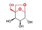
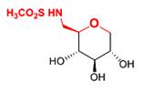 (1) (2)
Fig. 1. Thiosugar - 1,5-anhydro-6-deoxy-6-methane-sulfamido-D-glucitol (1) and chemical precursor 1,6-anhydro-5-C-hydroxymethyl-α-L-altro-pyranose(2).
In this work we compared cytotoxic and genotoxic effects induced by the studied chemicals in human colon adenocarcinoma cell line (LoVo), human lung adenocarcinoma epithelial cell line (A549) and chronic myelogenous leukemia (K562). A 12-h exposure of compounds hasn’t induced a significant decrease in the viability of LoVo and K562 cells. Only thiosugar increased the viability of A549 cells by 50% at 4 mM concentration. Both compounds evoked DNA damage in all studied cancer cells, as evaluated by the alkaline comet assay. Results from the neutral comet assay showed that both chemicals induced DNA double strand breaks (DSBs) and this was confirmed by the plasmid relaxation assay. DNA damage induced by thiosugar and its precursor was persistent and wasn’t removed during a 120-min repair incubation.
The results showed that thiosugar and its precursor induced DNA lesions in cancer cells. These compounds may be used in anticancer strategy in combination with DNA repair inhibitors.
This work was supported by the The National Science Centre, decision nr DEC-2011/01/B/NZ4/03391.
117. Biocompatibility of nanoparticles loaded
with photosensibilizers for practical use in support
of anticancer therapy
Jadwiga Pietkiewicz1, Urszula Bazylińska2, Joanna Rossowska3, Anna Choromańska1, Andrzej Gamian1,4, Kazimiera Anna Wilk2
1Department of Medical Biochemistry, Medical University of Wroclaw, Chalubinskiego 10, 50-368 Wroclaw; 2Faculty of Chemistry, Wroclaw University of Technology, Wybrzeze Wyspianskiego 27, 50-370 Wrocław; 3Institute of Immunology and Experimental Therapy, Polish Academy of Sciences, Weigla 12, 53-114 Wroclaw; 4Wroclaw Research Center EIT+, Stablowicka 147, 50-066 Wroclaw, Poland
In the recent years much effort has been made in the nanomedicine research to develop new effective and biocompatible drug delivery systems having a colloidal phase as a key prerequisite for their effectiveness. Among these systems an important role is played by the template-mediated nanocarriers that form high kinetically stable oil-core nanodispersions of enhanced therapeutic efficiency. The attractiveness of polymeric nanocarriers lies in their ability to incorporate many hydrophobic drugs into the oil phase thereby enhancing their solubility and permeation [1]. Our previous works has been carried out to explore the biological potential of the polymeric nanocapsules loaded with hydrophobic cyanines as effective delivery system of photosensitizer in photodynamic therapy of cancer cells [2,3]. In this report we describe physicochemical properties and biocompatibility of multilayer nanocapsules prepared by subsequent adsorption of opposite charged polyelectrolyte layers (dextran and chitosan sulfate sodium salt) on the nanoemulsion liquid core (layer-by-layer (LbL) approach) [1,3]. The L-b-L nanocapsules based on o/w nanoemulsion droplets stabilized by new generation of biocompatible saccharide-derived surfactants, i.e. linear 2-(dodecyldimethylammonio)-ethylgluco-heptonamide bromide (D2GHA-12) or dicephalic N,N-bis[3,3’(trimethylammonio)propyl] dodecanamide dimethylsulfate (C12(TAPAMS)2) and loaded with hydrophobic cyanine-type photosensibilizers: zinc phtalocyanine ZnPc or cyanine IR-780 were characterized for size, shape and morphology (by DLS, SEM and AFM) and colloidal stability. Additionally, analogs functionalized with polyoxyethylene glycol were prepared. The obtained nanocarriers offer simultaneously the ease of manufacture with little energy input, a high solubilization capacity and long time-stability. These nanocapsules were subjected to in vitro biological analysis for evaluate their biocompatibility and suitability for use in support of anticancer therapy. After parenteral administration, the nanocarriers may interact with plasma proteins and various cells present in the blood stream and for this reason therapeutic efficiency of delivered cargo may be diminished. We characterized biocompatibility of obtained nanocapsules by determination of cytotoxic effect on target cancer cells as well as by estimation of hemolytic activity against human erythrocytes. Additionally, we determined the cytotoxicity of nanocapsules against control normal cells (macrophages and endhotelial cells). Moreover, the degree of macrophage uptake of nanocapsules was examined and the serum albumin influence on this process has been establish for preliminary assessment of biodistribution. The low hemolytic potential and insignificant cytotoxicity against target cancer cells and macrophages as well as endothelial cells indicate good biocompatibility of obtained nanocapsules. Serum albumin at physiological concentration decreased macrophage uptake of nanocarriers, which suggests the prolongation of their half-life in the circulation. Nanocapsules functionalized with polyoxyethylene glycol were more safe for normal and cancer cells and do not undergo internalization by macrophages.
References:
[1] Bazylińska U., Skrzela R., Szczepanowicz K., Warszyński P., Wilk K.A. (2011) Soft Matter 7, 6113-6124.
[2] Wilk K.A., Zielińska K., Pietkiewicz J., Skołucka N., Choromańska A., Rossowska J., Garbiec A., Saczko J. (2012) Int. J. Oncol. 41, 105-116.
[3] Bazylinska U., Pietkiewicz J., Saczko J., Nattich-Rak M., Rossowska J., Garbiec A., Wilk K.A. (2012) Eur. J. Pharm. Sci. 47, 406–420.