


|
 |

Gliwickie Spotkania Naukowe 2007
Gliwice Scientific Meetings 2007
16-17 November
DISCOVERY AND VALIDATION OF BIOMARKERS BY MASS SPECTROMETRY
Tomasz Bieńkowski
Applera Poland
There is a critical lack of validated early biomarkers for most conditions and diseases.
Early diagnosis does enable treatment of less severe disease states, the use of less invasive techniques
and could potentially reduce the costs of healthcare systems.
Biomarker discovery and
verification/validation are two distinct workflows. During the discovery phase, a relatively small number
of samples with a high number of potential biomarker candidates are screened. The high-throughput provided
by the iTRAQTM reagent strategy together with 4800 MALDI TOF/TOF coverage allows for simultaneous
analysis of such samples. Once biomarker candidates have been identified with initial statistical
significance, these have to be validated. This validation workflow involves analyzing a large number of
samples with a relatively small number of candidates to establish the biological significance of the
biomarker candidates. Rather than switching to immunological techniques for this validation step, we
suggest a mass spectrometry based approach. This orthogonal strategy is a novel targeted, high throughput
quantitative multiplexed multiple reaction monitoring (MRM) approach. The approach relies on assay
development using a combination of MRMs to target specific peptides identified in discovery, followed by
MS/MS to confirm that the quantitative MRM signal results from the target peptide. This unique
verification workflow could be done on QTRAP like instruments.
The explanation why coupling LC with
MALDI TOF/TOF instrument is so useful and why QTRAP technology is needed to the verification step will be
presented.
MECHANISMS INTEGRATING SIGNALLING FROM G PROTEIN-COUPLED RECEPTORS AND EGF
RECEPTORS IN NORMAL AND MALIGNANT GASTROINTESTINAL CELLS
Thoralf Christoffersen, Olav Dajani,
Kristin Meisdalen, Monica Aasrum, Ingun H Tveteraas, Tormod Guren Dagny Sandnes
Department of Pharmacology, Faculty of Medicine, University of Oslo, Norway
We and others have found that several
agonists of heptahelical (G protein-coupled) receptors (GPCRs) may both stimulate and inhibit cell
proliferation, depending on the biological context. In many cells, notably glandular and lining epithelium
of the gastrointestinal tract, GPCR agonists, including prostaglandins (PGs), enhance cell growth by
acting in synergism with receptor tyrosine kinases (RTKs), particularly the epidermal growth factor (EGF)
receptor (EGFR). This is part of normal epithelial regulation and may also be important for tumour growth.
Insights into the underlying mechanisms are of biological interest and also may have therapeutic
implications, particularly in terms of strategies for combination therapy.
We have found that in the
hepatocarcinoma cell line MH1C1, prostaglandin E2 (PGE2) elicits EGFR phosphorylation, and induces
activation of the ERK-type of MAP kinases that is completely blocked by EGFR tyrosine kinase inhibitors
(gefitinib or AG1478), suggesting that PGE2 produces EGFR transactivation in these transformed cells.
Preliminary results indicated that this effect is mediated by Src. Furthermore, in the colon cancer cell
line HCT 116, stimulation with neurotensin (which is also a GPCR agonist) induced EGFR phosphorylation and
EGFR-dependent activation of PI3 kinase. However, the data indicated that in these cells,
neurotensin-induced activation of ERK, unlike PI3 kinase, did not involve EGFR transactivation.
In normal hepatocytes, where many GPCR agonists exert comitogenic effects, we found that neither early
response gene expression nor activation of DNA synthesis in response to GPCR agonists required EGF
receptor transactivation. Studies focusing particularly on the effect of PGE2, showed no evidence of EGFR
phosphorylation, and PGE2-stimulated activation of the ERK1/2 was totally insensitive to inhibitors of the
EGF receptor tyrosine kinase, suggesting that effects mediated via the PG receptor(s) do not involve EGF
receptor transactivation. However, PGE2 pretreatment, through a pertussis toxin-sensitive mechanism,
increased the magnitude of subsequent EGF-stimulated phosphorylation of Akt and markedly extended the
duration of EGF-induced ERK phosphorylation and ERK kinase activity. Thus, in these cells, PGE2 does not
act by transactivating the EGF receptor, but instead induces effects that are, at least in part, Gi
protein-mediated and result in a synergistic modulation of mitogenic signaling downstream of the EGF
receptor, including upregulation of the PI3K/Akt and the Ras/ERK pathways.
In conclusion, we have
identified two entirely different patterns of interaction between signalling pathways from receptors of
the GPCR and RTK families, where growth-promoting effects mediated via GPCRs may either occur via the EGFR
or converge with signalling downstream of the EGFR.
VOLTAGE-GATED Na+ CHANNEL UPREGULATION
AND POTENTIATION OF CELLULAR BEHAVIOURS IN METASTATIC DISEASE
Mustafa B. A. Djamgoz
Division of Cell Molecular Biology, Neuroscience Solutions to Cancer Research Group,
Imperial College London, South Kensington Campus, London SW7 2AZ, UK m.djamgoz@imperial.ac.uk
In an
electro-physiological approach to understanding the pathophysiology of metastatic disease, we have found
that strongly metastatic cells express voltage-gated Na channels (VGSCs) in neonatal splice forms. Most
work has been done on breast cancer (BCa) and prostate cancer (PCa) where VGSC activity potentiates a
range of metastatic cell behaviours, including "motility" (Fraser et al.,2003, 2005; Brackenbury et
al.,2007). This presentation will highlight a number of aspects of cancer cell motility in relation to
VGSC control, as follows:
Types of motility. Cellular motility has been measured and quantified in a
number of ways: a) 'wound-heal' assays [cf. early, lateral local motile activity]; (b) 'transverse
migration' [cf. intra/extravasation]; and (c) 'galvanotaxis' [cf. (b) taking into account also local field
or trans-cellular voltage gradients]. All three types of cellular motility were suppressed significantly
by suppressing VGSC activity using the highly specific blocker, tetrodotoxin (TTX). In the case of
MDA-MB-231 BCa cells, even ~30% reduction in VGSC (neonatal Nav1.5) activity by siRNA completely removed
the VGSC-dependent enhancement of motility.
Mechanisms upstream of VGSC expression. Our data are
consistent with VGSC upregulation occurring when BCa and PCa becomes hormone-independent and switches to
dependence on growth factors. Two major growth factors were found to be involved in PCa: nerve growth
factor and epidermal growth factor (EGF). Although both potentiated motile activity, it was only EGF that
signalled through VGSCs. Interestingly, in media containing a high level of insulin, VGSC activity
suppressed motility, raising the possibility of cellular motility being under servo-like control.
Mechanisms downstream of functional VGSC expression / activity. We know much less about these. On the
whole, there are two sets of possibilities: a) protein-protein interactions and (b) enzyme activity
stimulated by VGSC-mediated Na+ influx and/or subsequent changes in intracellular Ca2+ or pH. As an example
of the latter, evidence will be presented for VGSC/Na+-dependent PKA activity.
References:
1.
Brackenbury WJ et al. (2007). The neonatal splice variant of Nav1.5 potentiates
in vitro invasive
behaviour of MDA-MB-231 human breast cancer cells. Breast Cancer Res Treat. 101 : 149-160.
2. Fraser
SP et al. (2005). Voltage-gated sodium channel expression and potentiation
of human breast cancer
metastasis. Clin Cancer Res. 11 : 5381-5389.
3. Fraser SP et al. (2003). Contribution of functional
voltage-gated Na+ channel expression to cell behaviours involved in the metastatic cascade in rat prostate
cancer. I Lateral motility. J Cell Physiol 195: 479-487.
ION CHANNELS AND GENETIC DISORDERS THE CYSTIC FIBROSIS CASE
Krzysztof Dołowy
Department of Biophysics, Warsaw University of Life Sciences, Poland
The defect in single gene encoding chloride channel (CFTR) protein in epithelia
causes the most common fatal disease - cystic fibrosis. The CFTR protein is localized at the apical cell
surface and is activated by cAMP. The efflux of chloride ions and electrogenic sodium ion flow via
paracellular way causes water osmotic transport across the epithelia. A characteristic feature of the
disease is impaired chloride transport across several tissues including those of the airway epithelia. The
altered thicker mucus of greater viscosity leads to recurrent episodes of infections, inflammation and
destruction of affected organs. The origin of the most common mutation in CFTR gene occurred in Northern
Europe approximately in VIIth century AD. Its spread is owed to the selective advantage conferred to
heterozygous individuals against cholera and enterotoxic diarrhoeal diseases. There are three possible
ways to cure cystic fibrosis: gene therapy, activation of defective CFTR channel or activation of other
channel which will take over the CFTR function. None was proven to be effective yet.
MONTE CARLO METHODS FOR GENOMIC DATA ANALYSIS
Krzysztof Fujarewicz
Silesian University of Technology,
Gliwice, Poland;
krzysztof.fujarewicz@polsl.pl
DNA microarrays became a very popular method of gene
expression analysis. They can yield information about expression levels for almost the whole genome. The
data coming from DNA microarray experiments have the property that distinguishes them from data sets
obtained using other biometric techniques. In the case of microarrays the number of genes (using the
language of machine learning theory: features or attributes) is much greater than the number of
microarrays (observations). This property causes several problems with applying classical methods of
statistical analysis which may be applied improperly or results of these analyses may be over-interpreted.
For example, a frequently committed mistake is to select discriminating genes based only on p-value.
Moreover, most of classical statistical methods work under some assumptions which are not necessarily
fulfilled by microarray data.
In this presentation we show results of applying several Monte Carlo
methods such as: permutation tests and resampling methods. These methods, unlike classical statistical
methods, require weaker assumptions of applicability. They need more computational effort but, nowadays,
powerful computers are able to perform computations even on such huge data sets.
In the presentation
we show our two recently published methods based on bootstrapping: Bootstrap Based Feature Ranking (BBFR)
and Bootstrap Based Outlier Detection (BBOD).
We present the results of analysis for thyroid cancer
data set and for Barrett's Esophagus data set.
This study was supported by the Ministry of Science
and Higher Education, Poland, Grant No. PBZ-MNiI-2/1/2005.
EXPLORATIONS INTO BIOCHEMICAL PATHWAYS
Johann Gasteiger1,2
1Computer-Chemie-Centrum University of Erlangen-Nuremberg, Naegelsbachstr. 25, 91052 Erlangen, Germany
http://www2.chemie.uni-erlangen.de;
2Molecular Networks GmbH, Henkestr. 91, 91052 Erlangen, Germany http://www.molecular-networks.de
Living species have to
survive in a hostile environment. In order to achieve this goal they have to run chemical reactions to
produce energy for maintaining a desired temperature and they have to metabolize nutrients to convert them
into metabolic building blocks and, eventually, biological macromolecules.
Biochemical processes in
living organisms are often represented by complicated two-dimensional networks. Finding the desired
information, and, in particular, perceiving relationships between individual reactions in such networks
can be quite difficult. In order to assist in this endeavor, we have stored the contents of the poster
"Biochemical Pathways" originally distributed by Boehringer Mannheim (now Roche) in a reaction database
and have enriched it with additional information. The database contains 1,500 structures and 2,200
reactions. Small as this database is, it nevertheless stores information on the most important reactions,
those that keep us alive.
Searches can now be performed for names, full structures and substructures,
reaction partners, enzymes and coenzymes, organisms, reaction centers, etc. By using a standard structure
format, other chemical databases and computer programs can be connected to this database. Furthermore,
connection to bioinformatics databases can be made through enzyme names and enzyme codes. [1]
As an
application, we have investigated the geometric and electronic requirements of enzyme reactions.
Three-dimensional models were automatically built by the 3D structure generator CORINA for all molecules
involved in biochemical pathways. This then allowed testing the transition state hypothesis, stating that
the role of an enzyme is primarily to stabilize the transition state of a reaction. This hypothesis was
tested with inhibitors of some enzyme reactions by superimposing them on the intermediates of enzyme
reactions by GAMMA, a program based on a genetic algorithm. [2] This allowed us to establish the geometric
requirements of those reactions.
In order to investigate the electronic requirements of enzyme
reactions, various physicochemical effects such as charge distribution as well as inductive, resonance,
and polarizability effects were calculated for the atoms and bonds of the reaction center, i.e. those
directly participating in the reaction. These values were then used to train a self-organizing (Kohonen)
neural network, clustering these reactions. These clusters, by and large, correspond to the classification
of enzymes by the EC code. However, sometimes differences are observed indicating deficiencies of the EC
classification and pointing out that the physicochemical descriptors show finer details of enzyme
reactions.
The individual reactions of the reaction database have been connected into a network of
biochemical pathways. Methods have been developed to search in such a network over many reaction steps.
This allows one to find all pathways between two compounds and, thus, pinpoint alternative pathways if one
reaction step is blocked such as through deficiencies in an enzyme or through the down-regulation of a
gene.
Thus, this database provides deeper insights into the mechanism of biochemical pathways and can
also be used for making inferences on the metabolism of compounds.
References:
1. M. Reitz, O.
Sacher, A. Tarkhov, D. Truembach, J. Gasteiger, Org. Biomol. Chem. 2004, 2, 3226-3237.
2. S. Handschuh,
M. Wagener, J. Gasteiger, J. Chem. Inf. Comput. Sci, 1998, 38, 220-232.
CHARACTERIZATION OF HOMOZYGOUS DELETIONS IN LARYNGEAL SQUAMOUS CELL CARCINOMA CELL LINES
Giefing Maciej1,2, Martin-Subero Jose Ignacio2, Kiwerska Katarzyna1, Jarmuż Małgorzata1, Grenman Reider3, Siebert Reiner2, Szyfter Krzysztof1
1Institute of Human Genetics, Polish Academy of Sciences, 60-479 Poznan, Poland;
2Institute of Human Genetics, University Hospital Schleswig-Holstein Campus Kiel, Christian-
Albrechts University 24105 Kiel, Germany;
3Department of Otorhinolaryngology - Head and Neck Surgery
and Department of Medical Biochemistry, Turku University Central Hospital and Turku University, P.O. Box
52, FIN-20521 Turku, Finland;
4Department of Otolaryngology, University of Medical Sciences, 60-355 Poznań, Poland
The majority of classical tumor suppressor genes like CDKN2A or RB1 were identified
by the delineation of bi-allelic losses called homozygous deletions. To systematically identify homozygous
deletions in laryngeal squamous cell carcinoma and to unravel novel putative tumor suppressor genes we
screened three laryngeal squamous cell carcinoma cell lines (LSCC) using array Comparative Genomic
Hybridization (array-CGH).
Out of 31 candidate regions for homozygous deletions identified by
array-CGH, 5 were verified further by PCR. Among others, these homozygous deletions affected the tumor
suppressor CDKN2A and the apoptosis-inducing STK17A gene. To assess the frequency of the identified
deletions we investigated the affected sites in 9 additional LSCC cell lines. In 5 out of the 9 cell lines
the CDKN2A gene was homozygously lost. Thus, CDKN2A was homozygously deleted in a total of 7 out of 12
cell lines. No other recurrent homozygous deletions were found.
In this study we showed homozygous
deletions as a frequent mechanism of CDKN2A inactivation. Moreover, we identified several other genes,
including the putative tumor suppressor STK17A, which may be inactivated by homozygous deletions and thus,
potentially implicated in laryngeal squamous cell carcinoma development.
METAGENOMICS PROVIDES NEW PERSPECTIVES ON HUMAN HEALTH
Adam Godzik
Joint Center for Structural Genomics, La Jolla, CA 92037, USA
Metagenomics (Environmental Genomics) is based on applying modern genomic techniques
(DNA sequencing and/or proteomics) to the study of genetic material from microbial communities directly in
their environment. It discovered an unexpected genetic and metabolic diversity of the microbial life. In
most studies, previously known microorganisms comprised less than 0.1% of what was seen. One of the most
interesting and diverse environment studied by metagenomics is ... humans! Human gastrointestinal tract or
skin turned out to be rich in previously unknown microbial species, forming complex communities that play
an integral role in human health and disease. Billions of benign microbes that constitute human
microbiome, help us to digest food, break down toxins and fight off disease-causing microbes and form
complex network of interactions with our body. Many human diseases, from cancer, diabetes, inflammation to
obesity, can be correlated with specific changes in human microbiome. We present preliminary analysis of
human gut microbiome by DNA sequencing and proteomics, focusing on novel functions and pathways.
SUGARS IN NEW DRUG DESIGN - A LESSON FROM NATURAL PRODUCTS WITH ANTICANCER ACTIVITY
Grzegorz Grynkiewicz
Pharmaceutical Research Institute, Rydygiera 8, 01-793 Warsaw, Poland
Approximately half of the existing drugs have been derived from (or inspired by) natural products, while
recently obtained large synthetic combinatorial libraries fail to deliver experimentally validated new
drug candidates. Secondary metabolites, successfully exploited in medicinal chemistry as pharmacological
models or drug leads, frequently contain in their structure a glycosidic element, seemingly indispensable
for their biological activity. At a dawn of glycobiology and glycomic era we learn to appreciate molecular
recognition mechanisms of carbohydrates on a biopolymer level (which govern majority of the vital cell
sociology phenomena), but we are still mystified by functions performed by a single monosaccharide moiety
in a low molecular weight ligand. Examples of the structure - function relationship of glycons will be
drawn from natural products' pool as well as from synthetic anticancer drugs. Carbohydrate scaffolds,
ADEPT constructs and other pro - drugs will be discussed. Based on our own experience with anthracycline
anticancer antibiotics and new synthetic flavonoid glycosides, 2-deoxypyranosides are singled out as a
class of reasonably available derivatives for useful modification of pharmacological properties of
structurally complex and multifunctional drug lead compounds. An example of cell growth phase-selective
switch in the mechanism of action upon isoflavone glycosylation illustrates this point. In conclusion, we
postulate that glycodiversification of drug leads by application of glycal chemistry [1,2] offers new
opportunities in drug design and discovery.
References:
1. G. Grynkiewicz, W. Szeja, Synthesis
of the sugar moieties, in: "Anthracyclines Chemistry and Biology"(ed. K. Krohn), Top. Curr. Chem.,
Springer-Verlag Berlin Heidelberg 2008
2. W. Priebe, I. Fokt, G. Grynkiewicz, Glycal derivatives,
synthesis and chemical reactions, in: "Glycoscience; Chemistry and Chemical Biology"2nd ed., (eds. B.
Fraser-Reid, K. Tatsuta, J. Thiem), Springer-Verlag Berlin Heidelberg New York 2008
The study
supported by the Ministry of Science and Higher Education, Poland, Grant No. PBZ-MIN-014/P05/2004
WOUND HEALING AS A DIFFUSIONAL SORPTION PROCESS
Zbigniew J. Grzywna, Krzysztof Małysiak, Monika Krasowska
Section of Physics and Applied Mathematics, Faculty of Chemistry, Silesian
University of Technology, 44-100 Gliwice, ks. M. Strzody 9, Poland
The dynamics of wound healing
as a differential sorption process is considered. The different operators i.e. Smoluchowski, quasilinear
parabolic, and hyperbolic are compared. The velocity of a front tissue in all cases is calculated and
analysed. Each case presents a solid and convincing mechanism of a process in question. Comparison with a
previous approach, namely through Fisher's equation, is also provided.
MOLECULAR PET IMAGING AND GENE EXPRESSION PROFILING OF MALIGNANT TUMORS
Daria Handkiewicz-Junak
MSC Memorial Cancer Centre and Institute of Oncology, Gliwice Branch, Poland
The characterization of human diseases by
their underlying molecular and genomic aberrations has been the hallmark of molecular medicine. From this,
molecular imaging has emerged as new discipline that aims to visually characterise normal and pathologic
processes at the cellular and molecular levels of living organism. Advances in molecular imaging can
provide earlier and more precise disease diagnosis, improve disease characterisation and assessment of
therapeutic response.
Three different non-invasive, in vivo imaging technologies have developed: 1)
MRI; 2) nuclear imaging (PET, SPECT); and 3) optical imaging of small animals. The main advantage of
nuclear imaging is high intrinsic sensitivity (within nanomolar to fentomolar quantities of radiolabelled
probes), unlimited depth penetration and relative ease of radiolabelling molecular probes.
Molecular
imaging strategies are classified as direct and indirect. Direct molecular imaging is as characterised by
direct and specific interaction of molecular probe with a target while in indirect imaging most often
reporter-transgene technology is used, which couples a reporter gene with a complementary reporter probe.
This second approach is wildly applied in monitoring of gene therapy.
Although tremendous advances have
been made in molecular imaging there is still a gap between a quick development of functional genomic and
efficiency of molecular imaging. Gene expression profiling has shown that doze to hundreds genes play a
pivotal role in human cancers. The steady development of technologies of non-invasive detection and
measurement gene expression may help to fill this gap.
CONTROLLING THE ASSEMBLY OF THE CELLULAR MICROENVIRONMENT AND APPLICATIONS IN TISSUE REGENERATION AND REPAIR
David J.S. Hulmes
Institut de Biologie et Chimie des Protéines, CNRS/Université de Lyon UMR5086, Lyon, France
It is becoming increasingly apparent that the structure and physical properties of the
extracellular matrix making up the local cellular microenvironment have important roles in cell behaviour.
On the other hand, cells control the assembly and remodelling of the microenvironment through production
of structural proteins and extracellular enzymes. Thus cell-matrix interactions represent a two-way
dialogue involving both structural cues and remote enzymatic control. We are interested in the molecular
mechanisms that control matrix assembly and in particular the role of tolloid proteinases and associated
proteins. Tolloid proteinases have several substrates including structural proteins, proenzymes, growth
factors and their antagonists. We have recently identified a novel substrate-specific mechanism of tolloid
proteinase regulation that provides a new target for controlling collagen assembly in fibrotic disorders
(Moali et al., 2005; Blanc et al., 2007). The mechanisms that control the assembly of highly organised
tissues such as corneal stroma, which consists of an orthogonal stack of keratocyte containing collagenous
lamellae, are poorly understood. Both cellular control and liquid crystalline self-assembly mechanisms
have been invoked. In order to recreate this organisation for applications in tissue engineering, we have
developed a novel procedure for building a corneal stromal scaffold by carrying out fibril formation in
the presence of strong magnetic fields. In culture, corneal keratocytes penetrate this scaffold and become
aligned along the direction of the collagen fibrils (Torbet et al., 2007). This is the first step towards
reconstructing human corneas with the required optical and mechanical properties for use as alternatives
to corneal grafting.
References:
1. Blanc G, Font B, Eichenberger D, Moreau C, Ricard-Blum S,
Hulmes DJS, Moali C (2007) Insights into how CUB domains can exert specific functions while sharing a
common fold: Conserved and specific features of the CUB1 domain contribute to the molecular basis of
procollagen C-proteinase enhancer-1 activity. J Biol Chem 282: 16924-16933.
2. Moali C, Font B,
Ruggiero F, Eichenberger D, Rousselle P, Francois V, Oldberg A, Bruckner-Tuderman L, Hulmes DJS (2005)
Substrate-specific modulation of a multisubstrate proteinase. C-terminal processing of fibrillar
procollagens is the only BMP-1-dependent activity to be enhanced by PCPE-1. J Biol Chem 280:
24188-24194.
3. Torbet J, Malbouyres M, Builles N, Justin V, Roulet M, Damour O, Oldberg A, Ruggiero F,
Hulmes DJS (2007) Orthogonal scaffold of magnetically aligned collagen lamellae for corneal stroma
reconstruction. Biomaterials 28: 4268-4276.
GENETIC ANTIMELANOMA VACCINES: PROMISES AND OBSTACLES
Dariusz Iżycki
University of Medical Sciences at GreatPoland Cancer Center, Poznań, Poland
Classical therapy of advanced melanoma is disappointing but important progress has
been made in the understanding of melanoma immunology and biology. Malignant melanoma is an immunogenic
tumor, and its spontaneous remissions are associated with activation of the immune system. Discovery of
this fact induced a rapid progress in the field of immunology of this cancer and in the development of
novel strategies of immunotherapy. Recent achievements in cancer immunology have helped clarify the role
of the most important players in the development of host anti-melanoma immune response (including
different T cell subsets and dendritic cells). Moreover, these research attempts have allowed for
development of numerous new diagnostic tools to supervise immune responses in vaccinated patients
(penta-and tertramers techniques, ELISPOT assays and T-cell receptor analysis). Melanoma vaccines aim to
stimulate host immune responses against the patient's own tumor. A large number of immunotherapies of
advanced melanoma patients have been already studied in small-scale phase I-II trials. Several approaches
have demonstrated high rate of immune response which, unfortunately, did not become translated into
clinical benefit of patients.
445 advanced melanoma patients were, or have been enrolled since 1995
into Phase I and Phase II studies of various Hyper IL-6 and GM-CSF gene-modified whole-cell melanoma
vaccines that have been performed in the Department of Cancer Immunology at the University of Medical
Sciences in Poznań, Poland. In the phase II trial a 60% response rate was achieved (CR - 15%, PR - 15%, SD
- 25%). The DFS of patients immunized after surgical removal of metastases has been extended from 7 to 24
months. In this lecture the author, basing on his 12-year experience in clinical studies of melanoma
vaccines, will discuss the clinical outcomes of antimelanoma immunotherapy, novel biological and molecular
targeted therapies and vaccine trials' methodology.
BIOPOLYESTERS AND THEIR SYNTHETIC ANALOGUES OF STRUCTURE CONTROLLED AT THE MOLECULAR LEVEL THAT ARE SUITABLE
FOR REGENERATIVE MEDICINE
Marek Kowalczuk
Polish Academy of Sciences, Centre of Polymer and Carbon Materials, M. Sklodowskiej-Curie 34, 41-819 Zabrze, Poland
Aliphatic polyesters are the most representative examples of biodegradable polymeric materials. Among them polyhydroxyalkanoates (PHA) constitute natural
polymers of the renewable origin. PHA and their synthetic analogues, as well as copolymers with
alpha-amino acids, become increasingly attractive due to their biodegradability and biocompatibility,
which are the key requirements for a material to be applied in regenerative medicine.
Structural
characterization of biomaterials to be used in regenerative medicine as scaffolds for cartilage and soft
tissue engineering is essential for the understanding of their properties and function as, e.g., an
attractive tool for preparing growth factors' delivery systems. Using mass spectrometry technique, molar
masses and structural details of mass-selected macromolecular ions can be determined, thus elucidating the
chemical nature of the polymer and its end groups [1-4].
Recent results pertaining to preparation and
molecular-level characterization (ESI-MS technique in particular) of selected biodegradable and
biocompatible polyesters, including synthetic analogues of biopolymers, will be
presented.
References:
1. G. Adamus, M. Kowalczuk, Rapid Commun., Mass Spectrom. 2000, 14,
195
2. A. H. Arkin, B. Hazer, G. Adamus, M. Kowalczuk, Z. Jedliński, R.W. Lenz Biomacromolecules, 2001,
2, 623
3. G. Adamus, M.S. Montaudo, G. Montaudo, M. Kowalczuk, Rapid Commun., Mass Spectrom. 2004, 18,
1436.
4. G. Adamus, P. Rizzarelli, M.S. Montaudo, M. Kowalczuk, G. Montaudo, Rapid Commun., Mass
Spectrom. 2006, 20, 804
THEORETICAL BACKGROUND AND UTILITY OF COMMONLY USED SEQUENCE MULTIPLE ALIGNMENT TOOLS
Jacek Leluk
Department of Molecular Biology, Faculty of Biological Sciences, University of Zielona Góra, Zielona Góra, Szafrana 1 (bld. A-8), Poland
The multiple sequence
alignment is a fundamental step of comparative protein and genomic studies. It is an important
intermediate data source leading to obtain consensus sequence defining the whole sequence family,
explaining the variability pathways, locating the structurally and functionally significant regions, and
many other results, not only limited to the primary structural level. It is obvious that value and
reliability of these results strongly depend on correct adjustment of the aligned sequences. The related
problem is an accurate location of gaps. Otherwise all subsequent results are doubtful.
There are a
number of algorithms for accomplishing the alignment procedure, which are implemented in many programs.
Most of them refer to stochastic matrices of the observed nucleotide/amino acid replacement frequency. A
tremendous number of applications comply with the Markovian model of mutational amino acid replacement.
This work demonstrates that the approaches based on Markovian model and applying stochastic matrices
such as PAM and BLOSUM are not suitable for interpreting the protein variability occurring in nature. They
do not reflect the natural mechanism of molecular micro- and macroevolution. The methods of gap location
and continuity establishment are also not justifiable for the homologous proteins. The proposed solution,
based on genetic semihomology approach, takes into account both levels (nucleotide and amino acid)
simultaneously, to reflect the natural evolutionary process (consisting of two components: mutational
variability and natural selection). It applies the three-dimensional diagram of genetic relationships
between amino acids instead of stochastic matrices of the replacement frequency. The problem of gap
location and gap continuity is also discussed.
Acknowledgement: This study was supported by The
Centre of Excellence for Applications in Biomolecular Modelling and Bioinformatics -
BioExplortorium.
AN APPROACH TO DRUG DEVELOPMENT IN AN ACADEMIC ENVIRONMENT
Timothy Madden
Director, Pharmaceutical Development Center, Department of Experimental Therapeutics,
University of Texas M. D. Anderson Cancer Center, Houston, TX, USA
The PDC was initially proposed
in 1998 as a unique program to facilitate and optimize anticancer drug development within The University
of Texas M.D. Anderson Cancer Center (MDACC) from early stages of development through to FDA approval of
Investigational New Drug applications (INDs). This included relevant aspects of production, testing and
manufacturing of drug entities, as well as the clinical testing of such agents in relevant Phase I/II
clinical trials. The PDC was built around institutional core strengths in pharmacy, chemistry,
pharmacology, toxicology, veterinary medicine and other components of drug development in place within the
institution. Now with a continual pipeline of potential therapeutics under evaluation, and several agents
brought to early phases of clinical testing, the PDC has fulfilled its initial promise of being able to
facilitate development of novel therapeutic entities for the benefit of our patients.
WP744/RTA 744, a
novel anthracycline, which crosses the blood-brain barrier is one example of the strength of the PDC. This
innovative new DNA-binding agent was designed for the treatment of glioblastoma and developed as a lead
compound by Prof. Waldemar Priebe and subsequently followed by preclinical studies at the PDC prior to and
following licensing by Reata Pharmaceuticals, Inc. of Dallas, Texas. Research performed at the PDC
provided 85% of the IND content filed with the FDA. That IND was approved in December, 2005 and the
initial Phase I trial, under the direction of Dr. Charles Conrad of the UTMDACC Department of
Neuro-Oncology, was recently completed. This trial has provided the required information concerning the
toxicity and appropriate dose of RTA 744 for Phase II trials. It has also provided 2 complete responses, 2
partial responses, and 5 minor responses. This degree of activity is unheard of in early clinical trials
of drugs directed at the treatment of brain tumors. The FDA had recently granted orphan drug status for
RTA 744, in large part due to data provided to Reata by the PDC. These data were generated through
Sponsored Research agreements between the PDC and Reata. This type of activity clearly demonstrates that
the PDC aids in the fulfillment of UTMDACC's mission to "Make Cancer History" while at the same time
protecting and enriching the value of the institution's intellectual property.
THE POTENTIAL OF ENDORADIOTHERAPY FOR THE DEVELOPMENT OF HIGHLY SPECIFIC CYTOSTATICS
Walter Mier
Department of Nuclear Medicine, University of Heidelberg, Germany
With the advances in
genomics, molecular biology including gene vector technologies today's molecular imaging modalities have
strongly been improved. The major progress is based on peptide and antibody targeting vectors. When
labeled with ß--emitting radioisotopes these agents are applicable for endoradiotherapy and exploit the
targeting potential for highly specific therapeutic applications. This novel class of pharmaceuticals
offers the potential to develop patient-specific therapies and might provide the means to go beyond the
possibilities of current chemotherapy and radiation therapy. The clinical potential of a new
radiopharmaceutical relies not only on the high receptor affinity and selectivity, high metabolic
stability, low non-specific uptake and high specific accumulation but also on favorable blood clearance
and excretion kinetics. The clearance kinetics and excretion routes of a radiopharmaceutical are crucially
important for high target-to-nontarget ratios both for imaging and therapeutic applications. For targeted
radionuclide therapy (endoradiotherapy) applied are radioactively labeled carrier molecules, such as
monoclonal antibodies that possess high specificity for target antigens on the surface of tumor cells. In
the first instance the radiopharmaceutical is labeled with a single photon- or positron-emitting isotope,
e.g. 68Ga. This allows precise determination of the tumor uptake with PET. After substitution of the
nuclide by a particle emitting isotope e.g. Y90 the identical pharmaceutical can be used for
endoradiotherapy. Several of these drugs such as 90Y-rituximab (Zevalin), 131I-tositumomab (Bexxar) and
the somatostatin receptor-binding 90Y-DOTATOC are nowadays successfully applied in oncological therapy.
Future generations of endoradiopharmaceuticals will address yet unknown targets which might be identified
by screening techniques such as ribosome and phage display peptide libraries.
HARNESSING LOW DOSE RADIATION RESPONSES FOR RADIATION PROTECTION
Carmel Mothersill
Medical Physics and Applied Radiation Sciences Dept, McMaster University, Hamilton, Ontario, Canada. Email
mothers@mcmaster.ca
The biological effects of low dose radiation exposure are the subject of
intense research at present because of the recent upsurge in medical exposures due to advances in
diagnostic imaging. It is estimated that the average dose to a North American has doubled in the last 10
years from approx 3mGy/year to more than 6mGy. Biological responses to low doses are now known to differ
from high-dose responses. Among the most studied mechanisms are bystander effects, adaptive responses,
genomic instability and low-dose hypersensitivity. All are induced at doses of concern in the environment
and the clinic. Unlike high dose responses, which result from direct or indirect damage to DNA resulting
from radiation-induced strand breaks, most low-dose responses are similar to stress or adaptive responses
and are active defense mechanisms resulting in the induction of pathways which at the level of the
organism and species are protective, although they can result in death of damaged cells. While the
existence of such pathways is not disputed, their relevance in vivo is disputed and the exact underlying
mechanisms are poorly understood. This paper will discuss recent advanced in low-dose radiation biology
and link the phenomena to discoveries in the fields of stress biology and chemical ecology. In all these
fields, stress caused by exposure to environmentally damaging agents, induces protective chemicals which
signal to cells or individuals the need to induce protective mechanisms. In the field of radiation
biology, such research is in its infancy but in chemical and stress biology fields, there is a huge
literature including methods for identification of stress signal molecules and investigation of their
modes of action at the physiological and pharmacological level. Harnessing of these signal molecules would
not only benefit radiation protection in the environment and workplace but could provide a new generation
of drugs for use in the protection of normal tissues from radiation damage during therapy or
imaging.
Methods used in these investigations include medium-transfer-style bystander experiments
designed to determine whether non-targeted mechanisms of radiation damage are involved in the mechanism.
Also we use a reporter system which always responds to a bystander signal if one is present. This allows
us to screen living tissue harvested from organisms for possible expression of bystander signals after
exposure to radiation. The ability to use tissues harvested from organisms allows us to monitor the
effects of various treatments given in vivo.
Results so far suggest that bystander signaling is highly
conserved and appears to be a stress response mediated by signal molecules of the size and chemical
properties associated with alarm signals in nature. It is mediated in our system by a calcium flux which
probably enables cellular responses to be activated. The dose needed to activate the response is very low
- approx 3mGy (Fig 1). Preliminary experiments have confirmed that serotonin, nicotine and glycine all
modulate or simulate bystander signaling. Fig 2 a and b show sample data for serotonin.
The ability of
these small simple molecules to alter radiation response by affecting non-targeted mechanisms means these
substances might have a role to play in radiation protection and also in the clinic.
PHARMACOKINETIC DESCRIPTION OF GABAERGIC CURRENT MODULATION BY BENZODIAZEPINES IN NEURONS
Jerzy W. Mozrzymas, Tomasz Wójtowicz, Michał Piast, Katarzyna Lebida, Paulina Wyrembek and Katarzyna
Mercik
Laboratory of Neuroscience, Department of Biophysics, Wrocław Medical University, Chałubińskiego 3, 50-368 Wroclaw, Poland
Benzodiazepines (BDZs) are known to increase the amplitude
and duration of GABAergic inhibitory postsynaptic currents (IPSCs). Moreover, at low [GABA], BDZs strongly
enhance GABAergic currents suggesting up regulation of agonist binding while their action on
conformational transitions of bound receptors (gating) is a matter of debate. The purpose of the lecture
is to present our study in which we have examined the impact of flurazepam and zolpidem on mIPSCs by
investigating their effects on GABAAR binding and gating and by considering specific conditions of
synaptic receptor activation. It is known that synaptically released agonist remains within the synaptic
cleft for less than 1 ms indicating that postsynaptic receptors are activated in very dynamic conditions.
To describe the GABAA receptor kinetics with resolution adequate to the time scale of synaptic
transmission, the applications of exogenous GABA were performed using the ultrafast perfusion system
(agonist application within 0.1 ms). Synaptic currents (IPSCs) and current responses to exogenous GABA
were recorded using the patch-clamp technique. Flurazepam and zolpidem enhanced the amplitude and
prolonged decay of mIPSCs. Both compounds strongly enhanced responses to low [GABA] but, surprisingly,
decreased the currents evoked by saturating or half-saturating [GABA]. Analysis of current responses to
ultrafast GABA applications indicated that these compounds enhanced binding and desensitization of GABAA
receptors. Flurazepam and zolpidem markedly prolonged deactivation of responses to low [GABA] but had
almost no effect on deactivation at saturating or half-saturating [GABA]. Moreover, at low [GABA],
flurazepam enhanced desensitization-deactivation coupling but zolpidem did not. Recordings of responses to
half-saturating [GABA] applications revealed that appropriate timing of agonist exposure was sufficient to
reproduce either decrease or enhancement of currents by flurazepam or zolpidem. Recordings of currents
mediated by recombinant ("synaptic") a1b2g2 receptors reproduced all major findings observed for neuronal
GABAARs. We conclude that extremely brief agonist transient renders IPSCs particularly sensitive to up
regulation of agonist binding by BDZs. Moreover, our data suggest that BDZ-induced prolongation of IPSCs
may be due to an enhancement of current evoked by GABA spilling over from synapse.
Supported by
Wellcome Trust International Senior Research Fellowship in Biomedical Science (grant no.
070231/Z/03/Z).
METABOLIC PATHWAYS IN HUMAN PROSTATE CANCER CELLS: REGULATION BY VOLTAGE-GATED
NA+ CHANNEL ACTIVITY
Maria E. Mycielska and Mustafa B. A. Djamgoz
Neuroscience Solutions to
Cancer Research Group, Division of Cell and Molecular Biology, Imperial College London, South Kensington
Campus, SW7 2AZ London, UK
Prostate is a unique organ that produces and releases large amounts of
citrate. Up to 180 mM citrate can be found in prostatic fluid (Kavanagh, 1994). This is necessary for
vitality and motility of sperm. Elevated citrate production in prostatic epithelial cells (PECs) is
possible due to the unusual regulation of mitochondrial aconitase (mACNT) by Zn2+ , testosterone and
prolactin. Importantly, the amount of citrate in prostate cancer (PCa) drops to the levels found normally
in blood (~ 200 µM) (Costello and Franklin, 2000). Citrate release in normal PECs occurs through a K+-dependent mechanism whilst PCa cells express an additional, Na+-dependent component designed primarily
for citrate uptake (Mycielska and Djamgoz, 2004; Mycielska et al., 2005). Citrate is a metabolic substrate
that can be used by mitochodria (as a Krebs' cycle intermediate) or in cytoplasm for fatty acid (FA)
synthesis. Increased FA synthesis is associated with cancer, in particular PCa, where fatty acid synthase
(FAS) is considered to be a metabolic oncogene. We found that the expressions of FAS and cytosolic
aconitase (cACNT), an enzyme involved in NADPH production necessary for FAS activity, were elevated in PCa
cells (PC-3M) compared with PECs (PNT2-C2). Furthermore, preincubation in extracellular citrate resulted
in significant enhancement of the metastatic cell behaviours (MCBs) of PC-3M but not PNT2-C2 cells
(Mycielska et al., 2006)
Strongly metastatic PCa (eg PC-3M) cells have been shown previously to
express functional voltage-gated Na+ channels (VGSCs) (Grimes et al.,1995; Laniado et al.,1997; Diss et
al.,2005). We have determined the effects of long-term (24 and 48 h) preincubation of PNT2-C2 and PC-3M
cells in tetrodotoxin (TTX), a highly specific blocker of VGSCs, on citrate uptake and associated
metabolic pathways. Treatment of PC-3M cells with TTX resulted in (1) significant decrease of Na+-dependent citrate uptake mechanism, (2) decrease of expression and activity of cACNT, and (3) decrease of
MCBs (adhesion, motility and endocytic membrane activity); there was no effect on FAS expression. Similar
treatment of PNT2-C2 cells with TTX had no effect on any of the parameters studied.
It is concluded
that the VGSCs activity has a significant role in the regulation of FA synthesis in PCa cells through
control of (i) expression of citrate supply (Na+-dependent uptake) mechanism and (ii) cACNT activity
necessary for FA synthesis. Both these effects would increase PCa cells' metastatic potential and further
support the notion that functional VGSC expression is an early event in PCa progression.
References:
1. Costello L. Franklin RB. (2000). Oncology. 59, 269-282.
2. Diss JK, Stewart
D, Pani F, Foster CS, Walker MM, Patel A Djamgoz MB. (2005) Prostate CancerProstatic Dis 8, 266-273
3.
Grimes JA, Fraser SP, Stephens GJ, Downing JE, Laniado ME, Foster CS, Abel PD Djamgoz MB. (1995). FEBS
Lett. 369, 290-294.
4. Kavanagh JP. (1994). Prostate. 24, 139-142.
5. Laniado ME, Lalani EN, Fraser
SP, Grimes JA, Bhangal G, Djamgoz MB Abel PD. (1997). Am J Pathol. 150, 1213-1221
6. Mycielska ME
Djamgoz MBA. (2004). J Physiol. 559, 821-833.
7. Mycielska ME, Palmer CP, Brackenbury WJ Djamgoz MBA.
(2005a). J Physiol. 563, 393-408.
8. Mycielska ME, Broke-Smith TP, Palmer CP, Beckerman R, Nastos T,
Erguler K Djamgoz MBA. (2006). Int J Biochem Cell Biol. 38, 1766-1777.
TARGETED CANCER THERAPIES USING DERIVATES OF NATURAL PRODUCTS- -BREVININ-2R AND APOPTIN AS EXAMPLES
Soumya Panigrahi1,3, Saeid Ghavami3, Iran Rashedi1,2, Sudharsana R. Ande1,2, Emilia Wiechec5,
Subbareddy Maddika4, Thomas Klonisch3, Marek Los1,2,3,6
1MICB, CancerCare Manitoba,
2Dept. Biochem. Med. Genetics,
3Dept. Human Anatomy Cell Sci., Dept. Physiology, Univ. Manitoba, Winnipeg, Canada;
4Department of Therapeutic Radiology, Yale School of Medicine, USA;
5Institute of Human Genetics, University of Aarhus, Aarhus, Denmark;
6BioApplications Enterprises, Manitoba, Canada
Targeted cancer therapies have been the "holy grail" of onco-therapy ever since
researchers and clinicians have started to understand principles governing oncogenesis. While a lot of
hope (and research funds) have been invested in recent years towards pharmaco-genetics, immuno-therapy and
gene therapy, screening programs, or sometimes pure luck, have revealed new promising substances which
(semi-)selectively kill cancer cells. Here we communicate our progress on the characterization of cancer
(semi-)selective properties of anuran defensin Brevinin-2R and a viral protein Apoptin.
Brevinin-2R is
a novel non-hemolytic defensin that was isolated from the skin of the frog Rana ridibunda. It exhibits
preferential cytotoxicity towards human and rodent malignant cells, as compared to primary cells including
peripheral blood mononuclear cells, T-cells, and human lung fibroblasts. Jurkat and MCF-7 cells
over-expressing Bcl2, and L929 and MCF-7 over-expressing a dominant-negative mutant of a pro-apoptotic
BNIP3 (ΔTM-BNIP3) were largely resistant towards Brevinin-2R treatment. The decrease in
mitochondrial membrane potential (ΔΨm), or total cellular ATP levels, and increased reactive
oxygen species (ROS) production, but not caspase activation or the release of Apoptosis Inducing Factor
(AIF) or endonuclease G (Endo G), were early indicators of Brevinin-2R triggered death. Brevinin-2R
interacts with both early and late endosomes. Lysosomal membrane permeabilization inhibitors and
inhibitors of cathepsin-B and cathepsin-L prevented Brevinin-2R-induced cell death. Autophagosomes have
been detected upon Brevinin-2R treatment. Our results show that Brevinin-2R activates the
lysosomal-mitochondrial death pathway, and involves autophagy-like cell death.
Apoptin is a 14 kDa
viral protein and known to induce apoptosis in a wide range of transformed but not in primary cells.
During the initial phase of our study an array-based analysis demonstrated that Apoptin interacts with the
SH3 domain of Abl and the oncogenic fusion protein Bcr-Abl(p210). Immuno-precipitation assays revealed
that Apoptin also strongly interacts with PI3-K and Akt, all known to be involved in the regulation of
cell proliferation and other metabolic processes. Using deletion mutants, we have mapped the critical
interaction domains within Apoptin and PI3-K. Our data indicate that apoptin "hijacks" cell proliferation
attempts and re-directs them toward induction of cell death.
APPLICATION OF THE GAUSSIAN MIXTURE MODEL TO PROTEOMIC MALDI-TOF MASS SPECTRA
A. Polański, J.Polańska
Faculty of Automatic Control, Electronics and Computer Scence, Silesian University of Technology, 44-100 Gliwice, Poland
We present a methodology of analyzing matrix-assisted laser desorption ionization time of
flight mass spectra (MALDI-ToF MS) based on the Gaussian mixture decomposition. Gaussian mixture model is
fitted to the data by maximizing the likelihood function by using a version of the expectation
maximization (EM) algorithm. We applied the method to the data from head and neck cancer patients and
healthy volunteers. MALDI ToF MS spectra obtained from blood plasma samples were analyzed using the
Gaussian mixture model. Differentiating components (cancer versus control) were searched by applying
statistical tests to variables given by weights of Gaussian components. Computations led to the detection
of 10-20 reliable differentiating components. The obtained differentiating components envelop regions on
the mass-to-charge (m/z) axis where there are most significant differences between cancer and control
samples spectra. Their m/z values can be further processed with the aim of drawing biological
conclusions.
This study was supported by Ministry of Science and Higher Education, Poland, Grant
No. PBZ-MNiI-2/1/2005.
THE DESIGN AND DISCOVERY OF HIV INTEGRASE INHIBITORS
Jarosław Polański
Department of Organic Chemistry, Institute of Chemistry, University of Silesia, Katowice,
Poland,
polanski@us.edu.pl;
http://uranos.cto.us.edu.pl/~zchorg/
Reverse transcriptase, protease
and integrase are three enzymes encoded by the HIV virus. About 20 drugs for HIV therapy are currently
available on the market. However, none of the approved drugs targets integrase. It is believed that such a
drug can significantly improve the therapy. A number of HIV integrase inhibitors have been described in
the literature. The so-called diketoacids (DKA), the compounds found by Merck among the large compound
library collected from more than 250000 molecules, accomplished an important breakthrough. However,
clinical assays of several DKAs including L-870810 were stopped recently due to their toxicity. Just
recently Merck reported a novel DKA modification, namely MK-0518 that is now in phase III clinical trial.
We designed and synthesized a series of novel integrase inhibitors based on quinoline scaffold. The
molecular factors limiting the activity of these compounds will be discussed. A series of similar
compounds with antiproliferative activity have also been designed and synthesized. The structure-activity
relationships for this series will also be presented.
DEVELOPMENT OF INTEGRATED APPROACH TO TARGET BRAIN TUMORS: MODULAR DESIGN OF BLOOD-BRAIN BARRIER (BBB) PENETRATING DNA BINDING AGENTS
Waldemar Priebe
The University of Texas M. D. Anderson Cancer Center, 1515 Holcombe Blvd. Houston,Texas 77030, USA
Malignant gliomas are devastating cancers, which due to infiltration
and location are difficult to treat; they also cause significant mortality in young populations.
Development of effective chemotherapeutic strategies has been limited in part by the inaccessibility of
the CNS to pharmacological intervention. As a possible target, topoisomerase II (topo II) overexpression
has been documented in human gliomas and correlated to poor survival, but no effective topo II poison
capable of reaching the target tissue after systemic administration has yet been
developed.
Immunohistochemical studies of primary and secondary glioblastomas and their astrocytic
precursor tumors have demonstrated that similar to topo II, ATP-binding cassette (ABC) transporters like
MRP1, LRP, and P-gp are overexpressed in glioblastomas. We hypothesized that the presence of ATP-binding
cassette (ABC) transporters in the blood-brain barrier (BBB) might, in part, be responsible for limiting
the CNS penetration of most anticancer drugs, while their presence in tumor tissue also confers resistance
to wide range of drugs at a cellular level.
To identify new agents effective in vivo against
glioblastomas, we have developed an innovative approach combining our modular design of DNA-binding agents
allowing for the creation of unique libraries of DNA binders and potential topo II poisons. By
systematically screening such libraries, we have identified highly apoptotic compounds that can circumvent
Pgp and MRP1-mediated resistance mechanisms, suggesting that such compounds will be potent cytotoxins
against gliomas, while possessing the ability to cross the BBB.
We prepared and screened a selected
library of over 400 DNA binding agents against a panel of cells overexpressing P-gp and MRP1, identifying
< 10 compounds possessing the necessary characteristics from which the compounds WP744 and WP769 were
selected for more detailed evaluation. Both compounds are structurally related to the well-known
anticancer drug doxorubicin (DOX), but they possess in vitro and in vivo properties that are very
different from those of DOX.
WP744 and WP769 are significantly more apoptotic than DOX against both
wild-type tumor cells and multidrug-resistant tumor cell lines with the MDR1 and MRP1 phenotypes. WP769 is
also a significantly more potent topo II poison than either DOX or WP744. Both WP744 and WP769 cross the
BBB, reaching CNS and tumor concentrations that exceed that of plasma. In vitro, both are effective at
nanomolar concentrations in inhibiting growth of the glioma cell lines U87MG, D54MG, and U251MG.
Because of their unique biological characteristics (potent topo II poisons, ability to cross BB
barrier, activity against multidrug resistant tumors), these agents are uniquely placed to become
effective therapeutic agents for the treatment of GBM.
WP744/RTA744 is currently in Phase I
clinical studies in humans and observed activity include complete, partial, and minor responses and stable
disease.
DETERMINISTIC MODEL OF P53|MDM2 SIGNALING PATHWAY
Krzysztof Puszyński1, Tomasz Lipniacki2
1Faculty of Automatic Control, Electronics and Computer Scence,
Silesian University of Technology, Gliwice, Poland;
2Institute of Fundamental Technological Research,
Polish Academy of Sciences, Swietokrzyska 21, 00-049 Warsaw, Poland.
P53 is a transcriptional
factor which in healthy cells remains at low level under the control of its inhibitor, mdm2. It becomes
activated (phosphorylated) in response to DNA damage. When activated and present in high concentration, it
induces the transcription of numerous genes involved in cell cycle arrest and DNA repair. If the last
fails, p53 final job is to trigger the cell-death program called apoptosis. In this work we propose a new
two-feedback model based on positive and negative feedback loops introduced by Ciliberto et al. [1]. Using
our model, we considered the role of the time delay in positive feedback loop, which was neglected by
Ciliberto et al. We show that time delay, changes dynamics of the system and is crucial for inducing
apoptosis when irreparable DNA damage occurs.
This study was supported by the Ministry of Science
and Higher Education, Poland, Grant No. PBZ-MNiI-2/1/2005.
THE NEW PHOTOSENSITIZERS AND THEIR APPLICATION IN PHOTODYNAMIC THERAPY
Alicja Ratuszna
A. Chełkowski Institute of Physics, University of Silesia, Katowice, Poland
Photodynamic therapy (PDT) is an established modality for
the treatment of solid tumors and other accessible lesions. Photosensitized reactions are dependent on the
generation of reactive oxygen species, in particular singlet oxygen, which accounts for the damaging
effects on biological macromolecules, such as membrane lipids and proteins. Therefore, compounds that have
a good 1O2 yield are used as photosensitizers. Compared to current treatments (e.g., surgery, radiation
therapy, and chemotherapy) PDT is also relatatively non-invasive, can be targeted accurately and repeated
doses can be given without total-dose limitations associated with radiotherapy, and the healing process
results in little or no scarring. Regardless of all these facts, PDT has not yet gained general clinical
acceptance. There is still a need for isomerically pure photosensitizers. Those which are currently
approved absorb light in the visible spectral regions below 700 nm, thus enabling access to deeper
residing tumors. There should be also enhancement in efficiency of singlet oxygen generation which would
allow reducing concentration of the photosensitizer necessary to treat tumors as well as increasing
biodistribution selectivity of the photosensitizer.
Our goal is to develop and synthesize novel
photosensitizers based structurally on the porphyrin or chlorin ring, with much better physicochemical
features and then test their feasibility in vitro and in vivo.
So far, several compounds have been
synthesized at the University of Silesia and characterized by HPLC, absorption, X-ray diffraction, IR and
X-ray photoemission spectroscopy [1]. Some of them which seemed to be the most interesting as potential
photosensitizers were selected for in vitro studies [2, 3]. The next step will involve therapeutic studies
of tumor-bearing mice. In vitro, we assessed dark and phototoxicity by MTS and clonogenic survival assays.
Intracellular biodistribution of each compound was studied by confocal microscopy. To detect the mode of
cell death, besides specific fluorescent dyes used to differentiate dying cells on the basis of staining
and morphological criteria flow cytometry analysis was performed. All the results are presented on the
poster: Szurko et. al.: Chlorin as potential photosensitizer for photodynamic therapy
(PDT).
References:
1. G. Kramer-Marek, A. Szurko, C. Serpa, L. Arnaut, P. Kuś, J. Habdas A.
Ratuszna - Spectroscopic studies and singlet oxygen quantum yields of synthetic porphyrin derivatives -
potential novel photodynamic therapy agents - Physica Medica XX, Supl. 1, 46-48 (2004)
2. G. Kramer -
Marek, C. Serpa, A. Szurko, M. Wideł, A. Sochanik, M. Śnietura, P. Kuś, R. Nunes, L. G. Arnaut A. Ratuszna
- Spectroscopic properties and photodynamic effects of new lipophilic porphyrin derivatives: efficacy,
localization and cell death pathways, - Journal of Photochemistry and Photobiology, B84, 1 - 14
(2006)
3. A. Szurko, G. Kramer - Marek, M. Wideł, A. Ratuszna, J. Habdas P. Kuś - Photodynamic effects
of two water soluble porphyrins evaluated on human malignant melanoma cells in vitro - Acta Biochimica
Polonica 50, 1165-1174 (2003)
ROLE OF DIFFERENT PROSTANOID RECEPTORS IN THE GROWTH STIMULATORY EFFECT OF PROSTAGLANDINS
Dagny Sandnes, Kristin Meisdalen, Olav Dajani Thoralf Christoffersen
Department of Pharmacology, Faculty of Medicine, University of Oslo, Norway
Prostaglandins
are locally produced agents that mediate their effects through interaction with G protein-coupled
receptors. Nonsteroidal anti-inflammatory agents, which prevent the generation of prostaglandins through
their inhibition of cyclooxygenases, appear to prevent tumor development. Thus, there has been much
interest in the effects of prostaglandins on tumor cell proliferation, apoptosis, migration, and
angiogenesis. The majority of studies suggest that these effects on tumor cells are mediated through
interaction with EP2 and EP4 receptors. Numerous mechanisms appear to be involved, including activation of
ß-catenin-stimulated gene transcription and transactivation of EGF (epidermal growth factor) receptors by
various mechanisms.
In primary cultures of rat hepatocytes, prostaglandins exert a small stimulatory
effect on DNA synthesis on their own, compared with the strong stimulation of DNA synthesis induced by
mitogens, such as as EGF, and they act mainly by enhancing the growth stimulatory effect of mitogens.
Therefore, they belong to the group of substances that are termed comitogens. Using different prostanoid
receptor agonists and antagonists, we have examined the receptors and signaling mechanisms involved in the
effects of prostaglandins in hepatocytes. Although hepatocytes express EP2 and EP4 receptors, we have
found no evidence of their involvement in growth stimulation in these cells. The growth stimulatory
effects of prostaglandins in the hepatocytes are mediated mainly by Gi-coupled EP3 receptors, with a minor
contribution from Gq-coupled FP receptors.
ONCOGENIC JAK/STAT SIGNALLING PATHWAYS AND DESIGN OF JAK-KINASE INHIBITORS
Piotr Setny1,2, Waldemar Priebe3, Bogdan Lesyng1
1CoE BioExploratorium, Faculty of Physics, University of Warsaw, Warsaw, Poland;
2ICM, University of Warsaw, Warsaw, Poland;
3The University of Texas, MD Anderson Cancer Center, Houston, TX, USA
Recently recognized oncogenic signalling pathways involve the Signal Transducer and Activator
of Transcription (STAT) proteins. Seven members: STAT1 - STAT4, STAT6, as well as closely related STAT5a
and STAT5b, are activated by Janus Kinases (JAKs). In diverse human cancers, such like: lymphomas,
leukemias, melanomas, breast cancers, ovarian cancers, lung cancers, pancreatic cancers or prostate
cancers, constitutive activation of STATs has been detected. For review see e.g. [1].
In our studies we
concentrate on JAK2/STAT3 and JAK3/STAT5 signalling pathways. One hypothesizes that inhibition of JAK2
and/or JAK3 decreases activation of the STAT proteins, and in consequence inhibits the downstream
signaling. We found that compounds which are based on a caffeic acid scaffold, and a number of their
derivatives, are effective inhibitors of these pathways. Modeling of these inhibitors has been reported
[2,3]. During this conference new results will be presented. Synthesis of the inhibitors is being
optimized and is the subject of separate presentations by W. Priebe and W. Szeja. Experimental studies of
the influence of these inhibitors on tumor cell lines along with some elements of the JAK/STAT kinetic
model can be found in [4] and [5], respectively.
References:
1. H. Yu and R. Jove, The Stats of
Cancer - New Molecular Targets Come of Age, Nature Reviews, Cancer, 4, 97-105 (2004)
2. W. Priebe, I.
Fokt, S. Szymanski, T. Madden, Jia-Ju Bao, B. Lesyng, Ch. A. Conrad, M. Kupferman, J. L. Abbruzzese, J. N.
Myers, Design, Synthesis and Structure-Activity Relationships of Novel, Jak2/STAT3 Signaling Inhibitors,
97th Annual Meeting of the American Association for Cancer Research, April 1-5, 2006, Washington DC,
Proceedings of the American Association for Cancer Research, LB 298, 2006
(http://www.abstractsonline.com/viewer/SearchResults.asp).
3. P.Setny, B.Lesyng, W.Priebe, Modeling of
Possible Binding Modes of Caffeic Acid Derivatives to JAK2 Kinase, 5th International Conference:
Inhibitors of Protein Kinases and Workshop Session: Novel Molecular Design and Simulation Methods, Warsaw,
Poland, June 23rd-27th, 2007, in Acta Biochim. Polon. 64-65, Suppl.3, 2007
4. K. Swiatek-Machado, A.
Adach, A. Ellert-Miklaszewska, W. Szeja, W. Priebe, B. Lesyng, and B. Kamińska, Synthesis and Evaluation
of Anti-tumor Activity of Novel Inhibitors of JAK2/ STAT3 Pathway in Glioma Cells, 5th International
Conference: Inhibitors of Protein Kinases and Workshop Session: Novel Molecular Design and Simulation
Methods, Warsaw, Poland, June 23rd-27th, 2007, in Acta Biochim. Polon. 38-38, Suppl.3, 2007
5. M.
Rybinski, A.Gambin, Details of the JAK-STAT Signaling Pathway Model, 5th International Conference:
Inhibitors of Protein Kinases and Workshop Session: Novel Molecular Design and Simulation Methods, Warsaw,
Poland, June 23rd-27th, 2007, in Acta Biochim. Polon. 63-64, Suppl.3, 2007
Acknowledgements: These
studies are supported by Ministry of Science and Higher Education (PBZ-MIN 014/P05/2004) and CoE
BioExploratorium, University of Warsaw.
NATURAL PRODUCTS AS RADIATION RESPONSE MODIFIERS
Colin Seymour and Carmel Mothersill
McMaster University, Canada, seymouc@mcmaster.ca
Protection of cells and organisms against low doses of radiation is a complex
issue which must be considered at the level of cells, tissues and organisms. "Protection" at one level,
for example, prevention of cell death, may be adverse at another level, if it allows a damaged cell to
survive and form a malignant tumour. Conversely, death of a cell carrying damage can be protective for the
organism if it eliminates a damaged cell. Thus, it is important to understand the mechanisms involved in
protection against radiation damage at several hierarchical levels.
The use of natural products as
radiation response modifiers is very attractive. Many of these compounds are readily available and their
function and pharmacology is well understood. Some derive from venoms or natural defenses and are
currently used in medicine, others include vitamins, antioxidants or cofactors, which are tried and tested
nutritional supplements.
Radiation effects may be targeted or untargeted. Radiation may interact
directly within a cell causing a direct DNA lesion or it may elicit a bystander response from the
irradiated cell. A bystander effect is produced when the irradiated cell apparently exhibits no damage
from the radiation, but passes on biochemical signals, which induce neighbouring cells to apoptose or
undergo a number of other responses usually associated with irradiation such as mutation induction,
transformation, induction of ROS responses etc. Effects induced in progeny of non-targeted cells in
receipt of bystander signals include genetic instability, mini and microsatellite mutations and
carcinogenesis. A key characteristic of these non-targeted effects is that they occur at very low acute
doses (of the order of 5mGy) and saturate so that effective prevention requires an agent which can
effectively shut off the mechanism. While the mechanism is not fully known, it is thought to involve
signals from irradiated cells communicating via membrane receptors, to induce stress. There is evidence in
vivo from bomb survivors of the persistence of these effects for 50 years. The instability consequent on
the process can predispose to later carcinogenic insult. At low radiation doses (as might be predicted
from a dirty bomb where widespread, disruptive low level contamination is a desired outcome) untargeted
effects may predominate in terms of long-term major human health effects.
Our hypothesis is that
chemicals derived from marine invertebrates will be useful in terms of modifying and negating any long
term health consequences. Sessile benthic invertebrates including marine tunicates, cnidarians, and
sponges in particular, have developed an array of structurally unique bioactive natural products, which
have been demonstrated to afford the producing organism a competitive advantage in ecosystems such as
tropical coral reefs, characterized by extreme resource limitations. In addition to limited resources,
environmental pressures such as predation, fouling, competition for space and exposure to ultraviolet
radiation drive the production of these chemicals. In addition to the variety of toxic compounds produced
as defensive agents, organisms use highly coloured pigments to protect against the high levels of UV
radiation in tropical coral reefs and pigments such as these are known radioprotectors in radioresistant
bacteria .
This paper will review the literature concerning known radiation response modification by
natural products, with particular reference to substances which modify low- dose effects and will present
new data concerning the effects of some marine substances derived from sponges which we have found to
sensitise cells to radiation. Drawing together the data in this area should permit some conclusions to be
drawn about the mechanisms operating at low doses which can be targeted for radiation protection. We will
also present new preliminary data which uses natural products derived from marine sponges. These products
have been shown to have very active radiobiological activity. The structures are shown
below
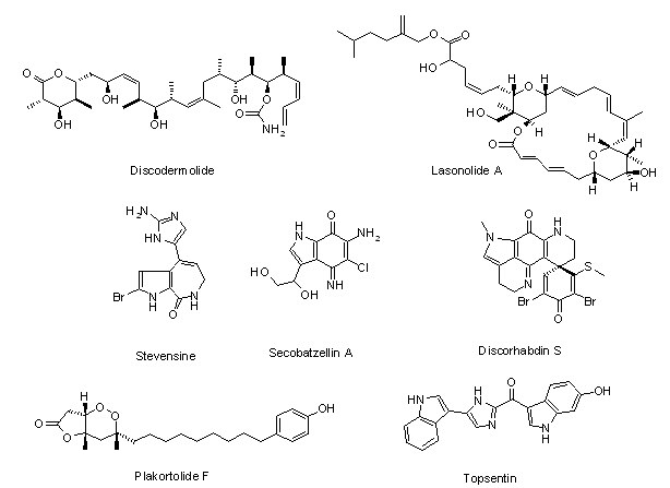
These compounds are radiation response
modifiers acting via bystander mechanisms.
GENETIC ENGINEERING OF ECM COMPOUNDS FOR REGENERATIVE MEDICINE
Aleksander L. Sieroń, Maciej Tarnowski, Anna Szydło
Department of General and
Molecular Biology and Genetics, Medical University of Silesia, Katowice, Poland, CoE for Study and
Teaching of Molecular Biology of Matrix and Nanotechnology, CoE Network BioMedTech
"Silesia"
Various human genetic disorders including mechanical damage leading to loss of tissue or
function of the organ need novel treatments. One possibility is genetic engineering. Two approaches are
presented, both related to collagen type I. Osteogenesis Imperfecta (OI) is a genetic disorder caused by
defects in COL1A1 or COL1A2 genes affecting production of procollagen type I and quality of its fibrils.
No cure for OI is available, except for orthopedic treatment and some prevention with orthopedic
equipment. A gene or stem cell therapy or combination of both could be the solution for OI. Here, we
present targeting col1a1 and col1a2 genes in rat mesenchymal stem cells with human homologous sequences.
Five hybrid DNA constructs comprising isogenous sequences of rat and human collagen genes were introduced
into the rat bone marrow stem cells. The G418 resistant clones were screened for human DNA. Over 90% of
resistant clones incorporated human DNA and 2% have had targeted endogenous collagen loci. Increasing
length of flanking sequences from 1 to 4kb increased 10-fold targeting loci genes. Also, DNA recombination
strategies for defining domains critical for collagen stability as well as for interactions with ECM
proteins, regulatory factors and cells are presented. Potential applications of results for tissue
engineering, also, are discussed. Our results open new possibilities of fixing collagen gene mutations in
patients, preferably using stem cells, expanding them and putting them back into the patient. Also, the
enrichment of novel collagens with specific domains will create better material for regenerative
medicine.
GLYCOCONJUGATES.DEVELOPMENT OF A NEW BIOLOGICALLY ACTIVE COMPOUNDS
Wiesław Szeja1, Tadeusz Bieg1, Anna Kasprzycka1, Gabriela Pastuch1, Ilona Wandzik1, Jadwiga Zawisza1, Grzegorz
Grynkiewicz2, Bogusław Szewczyk3
1Silesian University of Technology, 44-100 Gliwice, Poland;
2Pharmaceutical Research Institute, Rydygiera 8, 01-793 Warszawa, Poland;
3University of Gdańsk, Departament of Molecular Virusology, 80-822 Gdańsk, Poland
Carbohydrates are now recognized as
playing a significant role in numerous physiological responses [1].The molecular diversity of
carbohydrates offers a valuable tool for drug discovery[2].
Glycosyltransferases (GTS) are enzymes
responsible for processing of biomacromolecules. They regulate many cellular functions and are therefore
important targets in medicinal chemistry [3]. We report herein the synthesis of several analogues of
uridine, which were designed to act as inhibitors, through binding to the active site of the enzyme in
competition with natural donor substrates. In order to construct analogues of uridine diphospho sugars we
have chosen to exploit glycal chemistry. First, uridine and glycals were selectively protected.
Afterwards, addition of uridine derivative to a glycal, catalysed by triphenylphosphine hydrobromide, was
performed [4]. In this way we have synthesized, in a totally stereoselective manner and in high yields,
several uridine derivatives of 2-deoxy sugars.
In search for effective inhibitors of sugar-processing
enzymes the heteroaryl thioglycosides and products of their oxidation are substrate analogs. We present a
simple and efficient methodology for synthesizing glycosyl sulfoxides, inhibitors of GTS.
The effect of
inhibitors on penetration and propagation of swine fever virus (SFV) will be presented. Even when the
viability of SK6 cells was higher than 90%, low doses of this inhibitor (20 µg/ml), arrested the
propagation of CSFV virus and the viral yield was decreased by over 80%.
References:
1. Varki
A., Glycobiology 3(1993)97;.Dwek R.A, Chem.Rev., 96(1996)683
2. Gruner S.A.W., Locardi E., Lohof E.,
Kessler H., Chem. Rev.,102(2002)491
3. Witczak Z.J., Nieforth K.A., Eds., Carbohydrates in Drug Design,
Marcel Dekker
Inc., New York, 1997.
4. Jung K.H,. Schmidt R.R., Glycosyltransferase Inhibitors in
Carbohydrate-Based
Drug Discovery, C.-H.Wong (Ed) Viley-VCH, vol.2(2003) 609.
5. Bolitt Ch.,
Mioskowski S., Lee G., and. Falck J. R.., J. Org. Chem., 55, 5812 (1990).
COPOLYMERS OF SYNTHETIC BIODEGRADABLE SCAFFOLD WITH COLLAGEN TYPE I
Anna Szydlo1, Joanna Pogrzeba1, Maciej
Tarnowski1, Piotr Kurcok2, Michal Kawalec2, Michal Sobota2, Sieron Aleksander L.1
1General and
Molecular Biology and Genetics, Medical University of Silesia in Katowice, CoE for Study and Teaching of
Molecular Biology of Matrix and Nanotechnology, CoE Network BioMedTech "Silesia". Poland;
2Polish Academy
of Sciences, Centre of Polymer and Carbon Materials, Zabrze, CoE Polymers 2000, CoE Network BioMedTech
"Silesia", Poland
New biodegradable polymeric materials are needed for artificial ECM-like
scaffolds that mimic a three-dimensional environment in tissues. In this study we have focused on
production of such scaffolds using procollagen type I and polyhydroxybutyrate (PHB). We have tested their
feasibility for in vitro fibroblasts cultures. A higher number of cells were attached to plastic and PHB
coat. After 4 hours the number of cells on plastic surface was 182 cells/mm2 and on PHB itself the number
was 174 cells/mm2. After 8 hours fibroblasts attached to plastic numbered 192 cells/mm2 and on PHB itself
ithe count was 171 cells/mm2. The number of cells attached to Procollagen I and Procollagen I coating PHB
(Copolymer) was lower. On the other hand fibroblasts have spread much faster on the Copolymer and on
Procollagen I. Already at the 1st hour about 23% of cells did spread on Copolymer and on Procollagen I
itself, compared to only 1% spread on plastic and on PHB alone. After 2 hours of culture almost 80% of
cells did spread on both Procollagen I and on Copolymer, whereas, only 20% of cells have spread on plastic
and on PHB itself. Spreading on scaffolds was almost complete following 8 hours of culture, but not on
plastic and PHB. PHB itself showed to be a poor substrate for cell spreading but not for cell attachment.
Cell numbers spread on PHB itself were significantly lower than in cultures on plastic. Subsequently, we
have created a three-dimensional structure of PHB. The method of electrospinning was used to form
PHB-based fabric. The PHB fibers have created a 3D structure of the scaffold. Incubation of electrospun
disc of PHB with procollagen type I, in the MES buffer, allows copolymer formation. Scaffolds prepared as
described above were seeded with human fibroblasts. Attachment and spreading of fibroblasts was assessed
by AlamarBlue® dye assay. Our results indicate that copolymers of collagen I with PHB are useful for
building 3D scaffolds and could be successfully colonized with human fibroblasts. Such scaffolds could be
also utilized as assay systems to test different compounds in preclinical trials, as well as to create
prosthetics supporting cells in repairing damaged tissues and organs, e.g. skin grafts, tendons, etc.
Supporting grants: KBN2P05C02926, NN-1-015/05, RFSD Stipend Fund (EFS-2.6 ZPORR No.
Z/2.24/II/2.6/17/04 RFSD to AS 9/2005).
NOVEL TECHNOLOGIES IN THE STUDY OF IFN ALPHA ACTIVITY
Tokovenko B., Kuklin A., Perepelyuk M., Obolenskaya M.(obolenskaya@imbg.org.ua)
Institute of Molecular Biology and Genetics National Academy of
Sciences of Ukraine Zabolotnogo str. 150, 03143 Kyiv, Ukraine
Background: Interferon alpha
(IFNα) is used as main or adjuvant treatment in the therapy of viral infections and several types of
cancer. However, very little is known about the physiological effects of IFNα in the absence of viral
infection, and about the exact mechanisms of IFNα action. Although the list of IFNα direct and
indirect targets encompasses more than 300 genes the IFNα gene regulatory network has not been
defined.
The aim of the study was dual: a) to identify the genes of primary response to IFNα as
potential targets for further investigation and the initial information link in gene regulatory network
and b) to elucidate whether IFNalfa, a typical product of KC, and protein kinase R (PKR), classical
IFNalfa; target (both the components of innate immune response), are involved in liver regeneration.
Materials and methods: Identification of the genes of primary response to IFNα treatment was
conducted using bioinformatics search in the promoters of all rat protein-coding genes (as identified by
Ensembl) for the presence of evolutionary conserved interferon-stimulated response element (ISRE), the
binding site of the interferon stimulated gene factor 3 (ISGF3).
The rats after 2/3 partial hepatectomy
(PHE) and laparotomy (LAP) were used 1, 3, 6 and 12 h post-surgery to model correspondingly G0-->S
transition and acute phase response, the latter being a constituent part of the former. The gene
expression was assessed in liver samples, isolated KCs and hepatocytes by RT-PCR, antiviral test, Western
blot analysis and immunohistochemistry.
Results: Over 700 genes were identified to have
evolutionary-conserved ISRE sites in promoters, and thus are potential primary-response genes to IFNα
treatment.
PHE induces 2-fold increase of IFNalfa mRNA content and liver antiviral activity at 1h
post surgery, decrease to the values lower than control at 3h and normalization during 6 - 12 h period.
LAP induces half of IFNalfa mRNA content and the antiviral activity less than the detection limit. KCs
in both models are responsible for IFNalfa expression.
The level of PKR mRNA increases 2 times at
1h after PHE, decreases at 3h and again increases to the 1h-level during 6 - 12h period after PHE. The
PKR-antigen localizes in hepatocytes in the nuclei and cytoplasm with preferential localization in
cytoplasm at 6h post PHE. LAP induces early decrease of PKR mRNA level with its subsequent
up-regulation.
Conclusions: The genes of potential primary response to IFN alpha constitute the basis
for further elucidating the gene regulatory network induced by IFN alpha.
The changes in IFNalfa
and PKR expression may be essential for liver G0 -->S transition and acute phase response.
DNA REPAIR IN THE PATHOLOGY OF LUNG AND COLON CANCERS
Barbara Tudek
Institute of Biochemistry and Biophysics, Polish Academy of Sciences, Pawińskiego 5a, 02-106 Warsaw, Poland
Oxidative DNA
damage and its repair are involved in pathogenesis of human cancers. The main pathway of oxidative DNA
damage repair is base excision repair (BER). Functional studies performed on blood leukocytes have shown
that some BER pathways may be decreased in cancer patients and this may be one of risk factors for the
disease development. Decreased 8-oxoG excision rate was observed in lung as well as head and neck cancer
patients. Lung cancer patients revealed also lower rate of 1, N6-ethenoadenine (etaA) excision rate, DNA
damage induced by lipid peroxidation (LPO). Repair of another LPO-induced modification,
3,N4-ethenocytosine (etaC) was decreased only in individuals developing lung adenocarcinoma,
histologically the type of cancer with etiology linked to inflammations. Activity of BER proteins is
regulated by gene polymorphism, interaction of partner proteins and post-translational modifications.
Polymorphism of DNA glycosylases may change their enzymatic activity. Some of these polymorphisms increase
the risk of inflammation related cancers, e.g. colon, lung and other cancers. BER system efficiency may be
also regulated by reactive oxygen species and diet, which stimulate transcription of some DNA glycosylases
and the major human AP-endonuclease, APE1. The activity of repair enzymes may also be regulated by
carcinogenic process. Stimulation of BER genes transcription or deregulation of repair proteins activity
is frequently observed in cancer cells. Thus, modulation of BER proteins activity may be an important risk
factor for cancer development, and may also contribute to cancer progression.
CHARACTERIZATION OF NONADHERENT RAT BONE MARROW STEM CELLS (VSEL CELLS)
Ksymena Urbanek1, Halina Koryciak-Komarska1, Aleksander L. Sieroń1, Aleksandra Bryzek2, Magdalena Jackiewicz2, Piotr Czekaj2
1Department of
General and Molecular Biology and Genetics, Medical University of Silesia, Katowice, Poland, CoE for Study
and Teaching of Molecular Biology of Matrix and Nanotechnology, CoE Network BioMedTech "Silesia";
2Department of Histology, Medical University of Silesia, Katowice, Poland
Stem cells can
potentially offer treatment that many patients need. However, before using stem cells in the clinic, more
studies are required. Of greatest potential and plasticity are embryonic cells, but it is unlikely that
embryonic stem cell will be available at all for clinical use. Adult embryonic-like stem cells provide an
alternative, which is more ethically acceptable and which could be soon available for transplantation
purposes. Recent reports on cells with great plasticity, that were found in mice, claim that cells with
marker pattern Sca-1+-lin--CD45- are a population that could be used for clinical applications. Cells of
similar phenotype were also identified in human umbilical cord blood. They express antigens typical for
embryonic cells: SSEA-1, Oct-4, Nanog, Rex-1. The cells may also be present in other tissues of adult
individuals. However, little is understood so far about signals that induce stem cells to differentiate
into particular cell types, nor how to get grafted cells to integrate effectively into tissues and organs.
We postulate that these cells are present in bone marrow of adult rats and their differentiation depends
on Notch signaling involving four Notch receptors encoded by genes Notch 1, 2, 3, and 4 and ligands, Delta
1 and 2, and Jagged 1 and 2. This pathway is one of the basic signaling pathways involved in the
regulation of cell biology and metabolism in different physiological situations, both during embryogenesis
and organogenesis as well as in later life. Disturbances of this pathway have been already linked to
numerous pathological conditions, including cancer.
A START-TO-END APPROACH TO THE ANALYSIS OF PROTEOMIC DATA
Beata Walczak
Department of Chemometrics, Institute of Chemistry, University
of Silesia, Katowice, Poland
beata@us.edu.pl, www.chemometria.us.edu.pl
Comparative proteomics
focuses on the differences in the protein pattern from normal and pathological (or treated) samples. When
two-dimensional (2D) gel electrophoresis is used as an analytical tool for separating proteins, comparison
of samples is based on the resulting digital images of the 2D gel electropherograms. Thus, an overall
success of comparative proteomics critically depends on accuracy and reliability of the analysis of the
gel images. The available software deals differently with the main steps of data analysis, such as images'
preprocessing, images' warping, spot detection, etc. Different approaches cause a significant
software-induced variance [1] and often lead to a significant amount of missing data in the resulting
peaks' table [2].
In the proposed start-to-end approach these drawbacks are eliminated [3]. Namely,
the preprocessing step is optimized based on the classifier performance, analysis is performed on the
pixels instead of spots level, multivariate methods are used for model construction, and the proteins
differentiating the studied classes of objects are identified based on the embedded into the classifier
feature selection method, validated based on the Monte Carlo technique.
References:
1.
A.W.Wheelock, A.R.Buckpitt, Software-induced variance in two-dimensional gel electrophoresis image
analysis, Electrophoresis, 26 (2005) 4508-4520,
2. H.Grave et al.. Challenges related to analysis of
protein spot volumes from two-dimensional gel electrophoresis as revealed by replicate gels, Journal of
Proteome Research, 5 (2006) 3399-3410
3. M. Daszykowski, I. Stanimirova, A. Bodzon-Kulakowska, J.
Silberring, G. Lubec, B. Walczak, The start-to-end processing of two-dimensional gel electrophoretic
images, Journal of Chromatography A, 1158 (2007) 306-317
MAKING SENSE OF VOLTAGE SENSORS
Stephen H. White
Dept. of Physiology and Biophysics, University of California, Irvine, CA 92697-4560, USA
Voltage-gated ion channels open and close in response to changes in
transmembrane potential due to the motion of their voltage-sensor (VS) domains. Long known exclusively as
regulatory accessories for ion-channel pore domains, VS domains have now been identified in
voltage-activated phosphatases, and more recently a novel VS protein that functions as a voltage-gated
proton channel has been discovered. These findings and the crystallographic structure of the Kv1.2
potassium channel leave little doubt that the VS can exist as an independent transmembrane domain in lipid
bilayers. Given the long-standing assumption that charged amino acid side chains cannot be exposed
directly to hydrocarbon, how is it possible that VS proteins and their highly charged S4 segments can form
stable transmembrane structures? The answer is that the lipid bilayer is far more complex-and
interesting-than its usual lollypop cartoon suggests. Biological and physical evidence augmented with
molecular dynamics simulations will be presented that reveal the extreme adaptability of phospholipids
that arises from the privileged relationship between their phosphate groups and arginine residues. This
adaptability makes possible the independent stability of VS domains. [Research supported by the National
Institute of General Medical Sciences and the National Center for Research Resources.]
MASS SPECTROMETRY ANALYSES OF THE SERUM PROTEOME AN IMPORTANT TOOL OF CLINICAL PROTEOMICS
Piotr Widlak
Maria Sklodowska-Curie Memorial Cancer Center and Institute of Oncology, Branch in Gliwice;
15 Wybrzeze AK, 44-100 Gliwice, Poland
Analysis of the low-molecular-weight region of the blood
proteome (using either serum or plasma samples) is an emerging method of clinical proteomics. Such
analyses base on mass spectrometry (MS), on either relatively simple technologies like MALDI-TOF or more
sophisticated combinations of multidimensional chromatography and MS. Although no single peptide is
expected to be a reliable bio-marker in such analyses, multipeptide sets of markers selected in numerical
tests have been already shown in a few studies to have prognostic and predictive value in cancer
diagnostics.
We have initiated a large cohort study aimed to identify a set of polypeptide biomarkers
for early detection and monitoring therapy of breast cancer patients. In additional study we analyze serum
proteome of patients with head and neck cancer subjected to radiotherapy, aiming to identify biomarkers of
radiosensitivity. Low-molecular-weight polypeptides (2-10 kD) are analyzed using MALDI-TOF MS after
removal of albumin and other larger proteins from serum of cancer patients (in the course of therapy) and
matched healthy controls. Specific polypeptide patterns identified through mathematical analyses are
cross-correlated with clinical observations as well as results of routine biochemical and
histopathological analyses in an attempt to select reliable biomarkers that could be applied in cancer
diagnostics.
PROGNOSTIC MOLECULAR BIOLOGICAL MARKERS OF BREAST CANCER
Iosif Zalutsky, Raisa Smolyakova, Alexander Mashevsky, Alexander Dubrovsky, Maria Budko
N.N. Alexandrov Research Institute of Oncology and Medical Radiology, Minsk, Belarus
The principal prognostic factors of
breast cancer (BC) are lymph node metastases, the tumour size and spread, its grade, the status of
estrogen and progesterone receptors. At present, intensive search is being done in clinical oncology for
molecular biological markers of cellular origin, which would allow prognosticating the response to chemo-
and endocrine therapy, the disease course and long-term results of the treatment.
The molecular markers
include oncogenes and proto-oncogenes, oncoproteins, different growth factors and their receptors,
receptors of steroid and peptide hormones, suppressor genes and products of their expression,
hormone-dependent proteins, proteases participating in metastasizing processes, integrins responsible for
intercellular contacts. The determination of the tumour proliferative activity, of the hormonal status,
growth factor receptors in BC patients is of vital importance for selection of the specific treatment
regimen and prognosis of the disease course.
The objective of the study is to establish molecular
biological markers in BC patients.
The study material consisted of tumour tissue obtained from 60 BC
patients during surgical intervention. All the BC patients had histological diagnoses. Infiltrative ductal
carcinoma (45.1%) and infiltrative lobular carcinoma (37.1%) of differentiation grade 2 prevailed in this
group of BC patients. The age of the patients included in the study varied from 31 to 85 years.
To
define the expression of hormonal estrogen receptors (ER) and progesterone receptors (PR), Ki-67
proliferative protein, epidermal growth factor receptors (EGFR, Her-2/neu), p53 suppressor mutant protein,
bcl-2 oncoprotein, immunohistochemical technique was used with DAKO Cytomation (LSAB, En Vision) kits.
The results of our investigations demonstrate that 23 (39%) BC patients had estrogens-positive tumours and
24 (42.11%) were progesterone-positive. It was established that ER was most frequently found in
well-differentiated and moderately differentiated tumours while in high-grade BC the ER level was reduced.
The PR analysis detected its hyperexpression in well- and moderately-differentiated tumours. The presence
of steroid hormone receptors in BC is an indication of a relatively favourable prognosis and of potential
sensitivity of the tumour to endocrine therapy. Her-2/neu, a protooncogene- coding receptor 2 of human
epidermal growth factor, is a marker strongly influencing the antitumour treatment policy. Hyperexpression
of Her-2/neu oncoprotein was found in the tumour tissues of 24.6% of the patients. Her-2/neu
hyperexpression by the tumour is an independent marker of poor prognosis, high risk of recurrence and
decreased survival. The evaluation of Her-2/neu is of special prognostic significance in the cases of
small tumours. The expression of p53 suppressor mutant protein was detected in 57.1% of BC patients. Among
them slight positive staining occurred in 37.5% of the cases, and strong positive - in 59.4%. Positive
Ki-67 reaction was observed in 79.3% of tumour tissue specimens of BC patients. Among them, 41.4% of the
total number demonstrated high proliferative activity. Ki-67 index inversely correlated with tumour
differentiation grade (R= - 0.3024; p<0.05). High proliferative activity of the tumour in the BC patients
under investigation was associated with p53 expression (R=0.5259; p<0.05). At the same time, inverse
correlation with progesterone receptor expression was established (R= - 0.3129; p<0.05). Bcl-2 oncoprotein
hyperexpression was registered in 77.7% of cases. Proliferative activity of the tumour inversely
correlated with bcl-2 expression (R= - 0.3244; p<0.05). The evaluation of EGFR expression found 83.6% of
the specimens with negative EGFR reaction. EGFR hyperexpression was diagnosed in 6.6% of the BC patients.
In conclusion: determination of the prognostic markers allows to obtain an objective molecular
biological profile of the tumour and to shape the optimal antitumour treatment policy complying with
individual indications of BC patients.
|